Protoplast vs. Nuclei Isolation for scRNA-seq: A Comprehensive Guide to Methods, Applications, and Optimization
This article provides a detailed guide for researchers and drug development professionals on the critical sample preparation techniques of protoplast and nuclei isolation for single-cell RNA sequencing (scRNA-seq).
Protoplast vs. Nuclei Isolation for scRNA-seq: A Comprehensive Guide to Methods, Applications, and Optimization
Abstract
This article provides a detailed guide for researchers and drug development professionals on the critical sample preparation techniques of protoplast and nuclei isolation for single-cell RNA sequencing (scRNA-seq). It covers the foundational principles of how these methods enable cellular heterogeneity analysis in plants and animals, respectively. The content delivers step-by-step methodological protocols, addresses common troubleshooting and optimization challenges, and offers a direct comparative analysis to guide method selection for specific research goals, such as functional genomics in crops or cancer biology in humans. By synthesizing the latest advancements, this resource aims to equip scientists with the knowledge to generate high-quality single-cell data and accelerate discoveries in biomedicine and agriculture.
Why Sample Prep Matters: Unlocking Cellular Heterogeneity with Protoplast and Nuclei Isolation
The revolutionary power of single-cell RNA sequencing (scRNA-seq) to unravel cellular heterogeneity, discover novel cell types, and map developmental trajectories is fundamentally constrained by a critical initial step: the efficient and unbiased isolation of single cells or nuclei from complex tissues [1]. This process of tissue dissociation presents a universal challenge across biological research, as the quality of the resulting single-cell suspension directly dictates the resolution, accuracy, and reliability of all subsequent genomic data [2]. In plant systems, this challenge is compounded by the presence of a rigid cell wall, necessitating specialized approaches such as protoplast isolation or nuclei extraction to access the transcriptome [3] [4]. The choice between these two primary paths is not trivial; it represents a significant methodological branch point with profound implications for experimental outcomes. Protoplast isolation, which involves the enzymatic removal of the cell wall, can inadvertently induce wounding responses and transcriptional stress artifacts, potentially confounding biological interpretations [4] [5]. Conversely, single-nucleus RNA sequencing (snRNA-seq) bypasses the need for cell wall digestion by using isolated nuclei, thereby preserving the native state of many fragile cell types and enabling the use of frozen or archived tissues [4] [6]. This Application Note delineates detailed, optimized protocols for both routes, providing researchers with the tools to navigate the core challenge of sample preparation for high-fidelity single-cell transcriptomics.
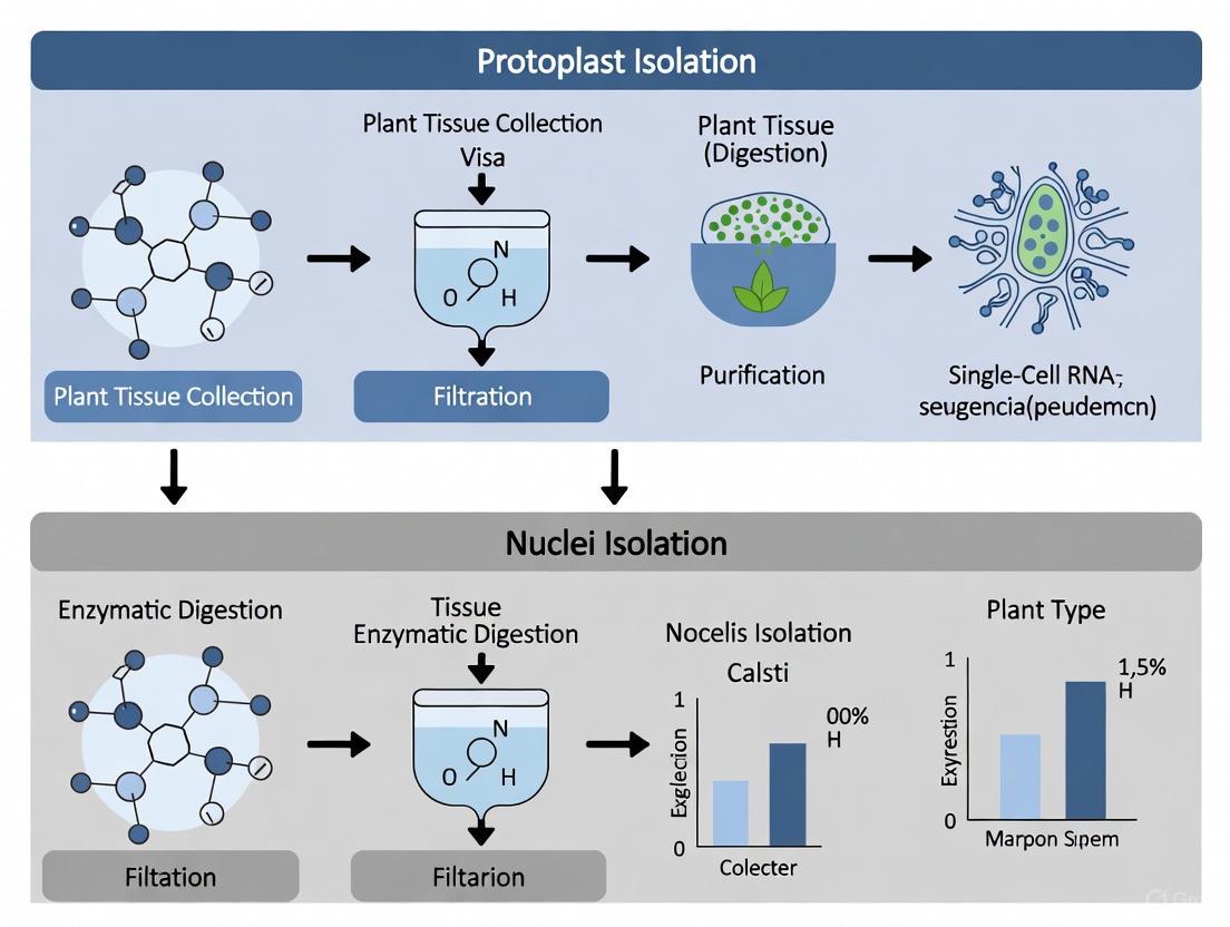
Methodological Approaches: Protoplast vs. Nuclei Isolation
Protoplast Isolation for Plant scRNA-seq
The isolation of protoplasts is a gateway to scRNA-seq for many plant species. A robust protocol, exemplified by work in cotton and Chirita pumila, hinges on precise enzymatic digestion and gentle handling to maximize yield and viability [7] [8].
Detailed Protocol for Cotton Root Protoplast Isolation [7]:
- Plant Material & Growth: Surface-sterilize cotton seeds (Gossypium hirsutum cv. ND601) and germinate on moist towels at 25°C for ~36 hours. Transfer seedlings with radicles ~1 cm long to a hydroponic system. Critical Tip: Use taproots from plants grown in hydroponics for 65-75 hours after germination for optimal results. Culture for less than 48 hours or more than 96 hours significantly reduces yield and quality.
- Tissue Preparation: Collect taproots from 25-50 seedlings. Using a sharp razor blade, cut roots into 0.5-1 mm thick translucent slices. Keep tissue immersed in solution to prevent desiccation.
- Enzymatic Digestion: Immerse tissue slices in 10 mL of freshly prepared enzyme solution. The solution consists of 1.5% (w/v) Cellulase R10, 0.75% (w/v) Macerozyme R10, 0.4 M mannitol, 20 mM KCl, 20 mM MES (pH 5.7), 10 mM CaCl2, and 0.1% BSA. Filter-sterilize before use.
- Incubation: Digest the tissue for 3 hours with gentle shaking (40-50 rpm) at 25°C in the dark. Monitor protoplast release microscopically.
- Protoplast Release & Filtration: Add an equal volume of W5 solution (154 mM NaCl, 125 mM CaCl2, 5 mM KCl, 2 mM MES pH 5.7) to the enzyme mixture and shake vigorously for 10 seconds. Filter the suspension sequentially through four layers of Miracloth and a pre-moistened 40 μm cell strainer into a round-bottomed tube. For scRNA-seq, a 30 μm strainer may be necessary to exclude larger cells.
- Purification & Storage: Centrifuge the filtrate at 100 x g for 5 minutes at 25°C using a swinging-bucket rotor with soft acceleration/deceleration settings. Gently discard the supernatant and resuspend the pellet in 5 mL of pre-chilled W5 solution. Keep the protoplast suspension on ice for 30-60 minutes before proceeding to counting, transfection, or scRNA-seq library preparation.
This protocol reliably yields up to 3.55 x 10^5 protoplasts per gram of tissue with a viability exceeding 93% [7]. A universal two-step digestion protocol developed for Chirita pumila further enhances viability (92.97%) and is applicable across diverse angiosperm organs [8].
Single-Nuclei Isolation for snRNA-seq
For tissues that are recalcitrant to protoplasting, prone to dissociation-induced stress, or uniquely archived, snRNA-seq offers a powerful alternative. The following protocol, adapted from human and plant studies, provides a general framework for nuclei isolation [9] [6].
Detailed Protocol for Single-Nuclei Isolation [9] [6]:
- Sample Preparation & QC (Basic Protocol 1 [6]): For frozen tissues, retrieve the sample on dry ice. Cut a small piece (e.g., ~5x5x5 mm) of the tissue of interest for RNA quality control. Isolate RNA and determine the RNA Integrity Number (RIN). A RIN of ≥5 is recommended to proceed with nuclei isolation, as it indicates sufficient RNA quality for downstream transcriptomics.
- Tissue Lysis/Homogenization: On ice, mince the main tissue sample to 0.5-1 mm pieces with a sterile scalpel. Transfer the pieces to a tube containing 1 mL of cold Lysis Buffer (1X PBS, 10 mM Tris-HCl pH 7.5, 0.0125% Triton X-100, 1 mM DTT, 0.2 U/μL RNase inhibitor). Critical Tip: Freshly prepare all reagents and keep them ice-cold. Decontaminate surfaces and treat equipment with RNase inhibitor to prevent RNA degradation.
- Mechanical Disruption: Place the tube on a magnetic stir plate with a micro stir-rod and incubate for 5 minutes at 100 RPM on ice. For tougher tissues, a mechanical homogenizer like a TissueLyser II may be required [6].
- Nuclei Recovery: Allow large debris to settle, then transfer the supernatant to a 15 mL tube containing 6 mL of cold Wash & Resuspension Buffer (1X PBS, 2% BSA, 0.2 U/μL RNase inhibitor). Repeat the lysis and wash steps 2-3 times to maximize nuclei yield.
- Purification & Filtration: Centrifuge the pooled supernatant at 600 x g for 5 minutes at 4°C. Gently resuspend the pellet in 1 mL of Wash Buffer and filter the suspension sequentially through 70 μm and 40 μm flow cytometer-compatible strainers.
- Quality Control: Centrifuge the filtered effluent again at 600 x g for 5 minutes and resuspend the final pellet in ~200 μL of Wash Buffer. Quantify nuclei using a hemocytometer or automated cell counter with DAPI staining. Assess nuclei integrity and the absence of contaminating cellular debris by microscopy.
This protocol is effective for obtaining intact nuclei with normal morphology and is compatible with a range of tissues, including clinical biopsies and various plant organs [9] [5].
Comparative Analysis: Protoplasts vs. Nuclei
Table 1: A comparative summary of protoplast-based scRNA-seq and nucleus-based snRNA-seq.
| Feature | Protoplast-based scRNA-seq | Nucleus-based snRNA-seq |
|---|---|---|
| Starting Material | Fresh, living tissue [3] | Fresh or frozen tissue [4] [6] |
| Key Challenge | Enzymatic digestion of cell wall; stress responses [4] [5] | Mechanical homogenization; RNA degradation [9] |
| Transcript Coverage | Whole-cell transcriptome (cytoplasmic + nuclear) [1] | Primarily nuclear transcriptome [4] |
| Cell Type Representation | May be biased against cell types with tough walls [4] | Broader representation, including large or fragile cells [6] |
| Typical Yield | High (e.g., 3.55 x 10^5/g in cotton roots) [7] | Variable, dependent on tissue and protocol [9] |
| Typical Viability | High (>90% with optimization) [7] [8] | Not applicable (assessed via nuclei integrity) |
| Best Suited For | Functional studies requiring living cells, transient expression [7] [8] | Complex, frozen, or difficult-to-dissociate tissues; archival samples [4] [5] |
The Scientist's Toolkit: Essential Reagents and Materials
Success in single-cell and single-nuclei workflows depends on a carefully selected set of reagents and tools. The following table lists key components for establishing these protocols.
Table 2: Key research reagent solutions and their applications in protoplast and nuclei isolation.
| Reagent / Material | Function / Application | Example Usage |
|---|---|---|
| Cellulase R10 / Macerozyme R10 | Enzymatic hydrolysis of cellulose and pectin in plant cell walls. | Protoplast isolation from cotton roots and Chirita pumila leaves [7] [8]. |
| Mannitol | Osmoticum to maintain pressure and prevent protoplast lysis. | Component of enzyme solution and washing buffers [7]. |
| Triton X-100 | Mild detergent for permeabilizing cell membranes to release nuclei. | Component of lysis buffer for nuclei isolation [9]. |
| RNase Inhibitor | Protects RNA from degradation during isolation procedures. | Essential addition to lysis and wash buffers for nuclei isolation [9]. |
| Polyethylene Glycol (PEG) | Facilitates plasmid DNA uptake into protoplasts for transfection. | PEG-CaCl2 mediated transformation in transient expression assays [8]. |
| DAPI (4',6-diamidino-2-phenylindole) | Fluorescent DNA stain for quantifying and assessing nuclei. | Staining for nuclei counting and viability assessment [9]. |
| FlowMi Cell Strainers (40 μm, 70 μm) | Removal of aggregates and large debris from nuclei suspensions. | Sequential filtration to purify single nuclei [9]. |
Workflow and Decision Framework
The decision to use protoplasts or nuclei for single-cell transcriptomics is multifaceted, depending on biological questions, sample availability, and technical constraints. The following workflow diagram maps this critical decision path and the subsequent experimental steps.
The journey from complex tissue to a high-quality single-cell suspension is the foundational step upon which the entire edifice of scRNA-seq is built. As this Application Note demonstrates, researchers are equipped with two powerful, complementary strategies: protoplast isolation and nuclei isolation. The choice between them should be guided by a careful consideration of the biological system, the experimental goals, and the practical constraints of sample availability. By adhering to the optimized protocols and decision framework outlined herein, scientists can reliably overcome the core challenge of sample preparation. This ensures that the resulting single-cell data truly reflects the underlying biology, free from the artifacts of preparation, thereby unlocking the full potential of single-cell transcriptomics to illuminate cellular heterogeneity and function in health, disease, and development.
Protoplasts, plant cells that have had their cell walls removed, serve as a fundamental resource in plant biotechnology and functional genomics. They provide a unique single-cell system ideal for a variety of applications, including transient gene expression, transformation, and the study of cellular processes. In the context of transcriptomics, protoplasts offer a pathway to single-cell RNA sequencing (scRNA-seq), enabling the resolution of cellular heterogeneity within complex plant tissues. However, the process of protoplast isolation induces rapid and significant transcriptional shifts, which can confound the analysis of native gene expression states. This application note details standardized protocols for protoplast isolation and introduces the alternative of single-nucleus RNA sequencing (snRNA-seq) for more accurate transcriptomic profiling, providing researchers with critical methodologies for accessing plant cellular transcriptomes.
Key Methodological Approaches for Transcriptomic Access
For transcriptomic studies, the choice between using protoplasts or isolated nuclei is critical. The table below summarizes the core characteristics of these two main approaches.
Table 1: Comparison of Transcriptomic Access Methods Using Protoplasts or Nuclei
| Feature | Protoplast-based scRNA-seq | Nuclei-based snRNA-seq |
|---|---|---|
| Biological Unit | Whole, living cell | Isolated nucleus |
| Transcriptomic Scope | Cytoplasmic & nuclear RNA | Primarily nuclear RNA |
| Tissue Starting State | Typically fresh tissue | Fresh or cryopreserved tissue [10] |
| Key Technical Challenge | Enzymatic cell wall digestion inducing stress responses | Mechanical homogenization to preserve nuclear integrity |
| Major Artifact Source | Transcriptional changes induced by prolonged enzymatic digestion [5] | Loss of cytoplasmic transcripts, ambient RNA |
| Ideal Application | Studies of cellular processes not affected by isolation stress | Profiling of complex tissues, archived samples, and immune responses [5] |
The Scientist's Toolkit: Essential Reagents for Protoplast Isolation
Successful protoplast isolation relies on a specific set of reagents. The following table lists key solutions and their functions in the protocol.
Table 2: Key Research Reagent Solutions for Protoplast Isolation
| Reagent / Solution | Function / Purpose |
|---|---|
| Enzyme Mixture (Cellulase, Macerozyme) | Digests cellulose and pectin in the plant cell wall [11] [12] [13] to release protoplasts. |
| Osmoticum (Mannitol or Sorbitol) | Maintains osmotic balance to prevent the fragile protoplasts from bursting [12]. |
| Plasmolysis Buffer | Often contains mannitol; initiates pre-plasmolysis of plant cells before enzymatic digestion, enhancing yield [11] [14]. |
| W5 Solution (or similar washing buffer) | Used for washing and purifying protoplasts after digestion; contains salts to maintain viability [11] [15] [13]. |
| MMg Solution | Contains mannitol and magnesium; used for resuspending protoplasts prior to transfection [15]. |
| Polyethylene Glycol (PEG) | Facilitates the delivery of DNA, RNA, or proteins (e.g., CRISPR/Cas9 RNP) into protoplasts [12] [14] [13]. |
Optimized Protocol for Mesophyll Protoplast Isolation
This protocol is adapted from established methods for leaf tissues from multiple species, including grapevine, pea, and Brassica carinata [11] [15] [13].
Materials and Reagents
- Plant Material: Young, fully expanded leaves from 3- to 4-week-old plants are optimal [11] [13]. For grapevine, 40 mg of young leaves is recommended [15].
- Enzyme Solution: Filter-sterilized solution containing:
- W5 Solution: 154 mM NaCl, 125 mM CaCl₂, 5 mM KCl, 2 mM MES, pH 5.7 [11] [13].
- Plasmolysis Solution: 0.4 M mannitol [11].
Step-by-Step Procedure
- Plant Growth and Leaf Preparation: Grow plants under controlled conditions. Excise young leaves and remove the midrib. Cut leaf tissue into thin strips (0.5–1.0 mm) using a sharp razor blade. The "strip-cutting" method is superior to random cutting for yield [15].
- Plasmolysis: Transfer the sliced tissue into a Petri dish containing Plasmolysis Solution. Incubate for 30 minutes in the dark at room temperature.
- Enzymatic Digestion: Replace the plasmolysis solution with the pre-cooled Enzyme Solution. Use approximately 10 mL of enzyme solution per gram of leaf tissue. Incubate in the dark for 6–16 hours at 23–25°C with gentle shaking (30–40 rpm).
- Protoplast Release and Purification:
- Gently swirl the digestion mixture to release the protoplasts.
- Add an equal volume of W5 solution to stop the enzymatic reaction.
- Filter the suspension through a 40-100 μm nylon mesh to remove undigested debris [11] [15].
- Transfer the filtrate to a centrifuge tube and spin at 100 × g for 5–10 minutes.
- Carefully remove the supernatant. Resuspend the pellet (protoplasts) in W5 solution.
- Repeat the washing step twice.
- Viability and Yield Assessment: Resuspend the final protoplast pellet in an appropriate volume of W5 or MMg solution. Determine protoplast density using a hemocytometer. Assess viability via staining (e.g., Fluorescein Diacetate, FDA) or by observing cytoplasmic streaming [14]. Yields can vary from 75 × 10⁶ protoplasts/g tissue in grapevine [15] to transfection efficiencies of up to 59% in pea [13].
An Alternative Pathway: Single-Nucleus RNA Sequencing (snRNA-seq)
To circumvent the transcriptional artifacts of protoplasting, snRNA-seq uses isolated nuclei. The workflow diagram below outlines the key steps for this method.
snRNA-seq Protocol for Cryopreserved Tissues
This protocol is adapted from a method demonstrating effective nuclei extraction from low-input (15 mg) cryopreserved human tissues, a approach that is directly transferable to plant research [10].
- Tissue Homogenization: Mince 15-30 mg of cryopreserved tissue on dry ice. Transfer to ice-cold lysis buffer (10 mM Tris-HCl pH 7.4, 10 mM NaCl, 3 mM MgCl₂, 0.05% NP-40). For plant tissues, the buffer may require optimization.
- Dounce Homogenization: Use a loose (A) and/or tight (B) Dounce homogenizer. The number of strokes (e.g., 10-20 strokes) must be empirically determined for each tissue type to balance nuclei yield and integrity [10] [16].
- Nuclei Purification: Filter the homogenate through a 30 μm strainer. Layer the filtrate over a 29% iodixanol cushion and centrifuge at 1000 × g for 10-20 minutes at 4°C. This step effectively pellets nuclei while separating them from cellular debris [10].
- Nuclei Sorting and QC: Resuspend the pellet in a nuclei washing buffer containing RNAse inhibitor. Stain nuclei with a viability dye like 7-AAD and sort using Fluorescence-Activated Cell Sorting (FACS) to select for intact, high-quality nuclei [10]. Confirm integrity and concentration by microscopy before proceeding to library construction.
Both protoplast isolation and nuclei isolation provide viable paths to transcriptomic access in plants. The choice of method hinges on the specific research question. Protoplasts are excellent for functional assays and transient transformation, but their use in scRNA-seq is compromised by isolation-induced stress responses. For high-fidelity transcriptomic mapping of complex plant tissues, especially when using cryopreserved samples or studying rapid responses like immunity, snRNA-seq from isolated nuclei offers a robust and artifact-minimized alternative. By applying the detailed protocols and considerations outlined in this document, researchers can make an informed choice and successfully implement these techniques in their investigations.
Single-nucleus RNA sequencing (snRNA-seq) has emerged as a powerful alternative to single-cell RNA sequencing (scRNA-seq), particularly when working with complex, tough, or cryopreserved tissues that pose significant challenges for traditional protoplast isolation methods. While scRNA-seq provides unprecedented insights into cellular heterogeneity, its application is limited by requirements for fresh tissues and the technical challenges of dissociating certain cell types without introducing artifacts. Nuclei isolation overcomes these limitations by enabling transcriptomic profiling of tissues that are difficult to dissociate, including frozen clinical samples, formalin-fixed paraffin-embedded (FFPE) archives, and complex organs like brain and adipose tissue [10] [17] [18].
The fundamental advantage of snRNA-seq lies in its compatibility with preserved tissues and its ability to minimize dissociation-induced transcriptional stress responses. Unlike whole-cell approaches that require immediate processing of fresh samples, nuclei can be isolated from tissues stored in biobanks, thereby unlocking vast repositories of clinical specimens for transcriptomic analysis [10]. This technical advancement is particularly valuable for studying human diseases, developmental processes, and cellular responses in contexts where fresh tissue procurement is impractical or impossible. Furthermore, snRNA-seq enables the profiling of cell types that are notoriously difficult to isolate intact, including neurons, adipocytes, and cardiomyocytes, thus providing a more comprehensive view of tissue heterogeneity [18].
Core Principles: Advantages of Nuclei Over Whole Cells
Technical and Biological Rationale
The transition from single-cell to single-nucleus RNA sequencing is motivated by several technical and biological considerations. First, nuclei maintain transcriptional profiles that closely correlate with those of whole cells, making them reliable proxies for cellular states [10]. Second, the nuclear membrane provides a protective barrier that preserves RNA integrity during the isolation process, reducing degradation artifacts that can compromise data quality. Third, nuclei are more resistant to mechanical stress than whole cells, allowing for more vigorous processing methods that ensure representative sampling of all cell types within a tissue [19].
Perhaps the most significant advantage of snRNA-seq is its compatibility with frozen and fixed specimens. Clinical samples are often cryopreserved or formalin-fixed to preserve morphology for pathological assessment, rendering them unsuitable for standard scRNA-seq protocols. snRNA-seq successfully bridges this gap, enabling transcriptomic analysis of samples that would otherwise be inaccessible for single-cell studies [10] [17]. This compatibility has profound implications for biomedical research, as it facilitates the analysis of well-annotated clinical cohorts with extensive follow-up data, thereby strengthening correlations between transcriptional signatures and clinical outcomes.
Applications Across Challenging Tissue Types
- Cryopreserved tissues: Enables analysis of rare clinical specimens with just 15 mg input material [10]
- FFPE archives: Unlocks decades of preserved clinical samples for transcriptomic studies [17]
- Complex organs: Maintains cellular diversity in brain, adipose, and other heterogeneous tissues [19] [18]
- Delicate cell types: Preserves transcriptional profiles of neurons, adipocytes, and other fragile cells [18]
Established Protocols and Methodologies
Versatile Low-Input Protocol for Cryopreserved Tissues
Principle: This method enables robust nuclei isolation from minimal amounts (15 mg) of cryopreserved human tissues, making it particularly valuable for rare clinical specimens [10].
Protocol Steps:
- Tissue Preparation: Cryopreserved samples are minced in a pre-cooled mortar on dry ice using a scalpel, then transferred to 15 mL tubes.
- Homogenization: Add 3 mL of ice-cold lysis buffer (10 mM Tris–HCl pH 7.4, 10 mM NaCl, 3 mM MgCl₂·6H₂O, 0.05% NP-40). Homogenize using a Dounce homogenizer with pestle selection (loose or tight) and stroke numbers optimized for specific tissues.
- Lysis Control: Incubate on ice for 5 minutes, then stop the reaction with 5 mL of ice-cold nuclei washing buffer (0.5X PBS, 5% BSA, 0.25% Glycerol, 40 units/mL Protector RNAse inhibitor).
- Filtration and Purification: Filter through 30 µm MACS strainers, then centrifuge for 10 minutes at 1000 g (4°C).
- Density Gradient: Resuspend pellets in nuclei washing buffer, then add 1 mL of 50% (wt/vol) iodixanol. Gently layer on top of 2 mL cushion of 29% (wt/vol) iodixanol.
- Nuclei Sorting: Stain with 7-AAD for 10 minutes, then sort using a BD FACSAria Fusion with 70 µm nozzle to collect intact nuclei [10].
Tissue-Specific Optimization:
- Brain tissue: Use pestle B (tight clearance) with 15 strokes
- Bladder tissue: Use pestle A (loose clearance) with 10 strokes
- Lung tissue: Use pestle B with 20 strokes
- Prostate tissue: Use pestle A with 15 strokes
snCED-seq: Advanced Method for FFPE Tissues
Principle: The Cryogenic Enzymatic Dissociation (CED) strategy enables high-yield nuclei extraction from FFPE samples by addressing the challenges of RNA cross-linking while preserving RNA integrity [17].
Protocol Steps:
- Deparaffinization: Hydrate FFPE sections using standard histology protocols.
- Protein Digestion: Incubate with proteinase K (optimized concentration) at low temperature (4°C) instead of conventional high-temperature incubation.
- Surfactant Treatment: Use sarcosyl as an anionic surfactant instead of SDS or Triton X-100 for better nuclear membrane preservation.
- Nuclei Release: Gentle mechanical agitation in cryogenic conditions to release nuclei without filtration or ultracentrifugation.
- Quality Assessment: Verify nuclei integrity and count using epifluorescence microscopy [17].
Key Advantages:
- 10x increase in nuclei yield compared to conventional methods
- Significant reduction in hands-on time
- Minimal secondary RNA degradation
- Preservation of intranuclear transcripts
- Enhanced gene detection sensitivity with lower mitochondrial and ribosomal contamination
Robust Protocol for RNA Quality Preservation
Principle: This method addresses tissue-specific variations in RNase activity that can compromise nuclear RNA quality, particularly in challenging tissues like adipose tissue [18].
Protocol Steps:
- RNase Inhibition: Incorporate vanadyl ribonucleoside complex (VRC) during nucleus isolation to maintain RNA quality across diverse tissue types.
- Tissue Homogenization: Process tissues in chilled homogenization buffer containing VRC and recombinant RNase inhibitors.
- Nuclei Purification: Centrifuge through appropriate density medium to separate intact nuclei from debris.
- Quality Control: Assess RNA integrity and nucleus morphology before proceeding to library preparation [18].
Performance: This optimized protocol successfully maintains nuclear RNA integrity in all tested tissues except pancreas and spleen, with well-dispersed nucleus populations without clumps. Nuclear RNA remains intact for up to 24 hours when stored at 4°C, significantly outperforming standard protocols where severe degradation occurs within 2 hours [18].
Comparative Analysis of Isolation Methods
Method Performance Across Tissue Types
Table 1: Comparison of Nuclei Isolation Methods and Their Applications
| Method | Input Requirements | Key Advantages | Tissue Compatibility | Nuclei Yield | RNA Quality |
|---|---|---|---|---|---|
| Low-Input Cryopreserved [10] | 15 mg cryopreserved tissue | Minimal input requirement, FACS sorting | Brain, bladder, lung, prostate | 1,550-7,468 nuclei per sample | High (comparable to fresh tissues) |
| snCED-seq [17] | Single 50 μm FFPE section | 10x higher yield, works with FFPE archives | Brain, liver, kidney, spleen, intestines | >1 million nuclei per gram tissue | Enhanced gene detection sensitivity |
| Sucrose Gradient Centrifugation [19] | ~30 mg fresh/frozen tissue | Well-established, cost-effective | Brain cortex | ~60,000 nuclei per mg input | Defined individual nuclei, minimal debris |
| Machine-Assisted Platform [19] | ~30 mg fresh/frozen tissue | Automated, minimal variability | Brain cortex | ~60,000 nuclei per mg input | Well-separated, intact nuclei (99% integrity) |
| Spin Column-Based [19] | ~30 mg fresh/frozen tissue | Fast processing, no special equipment | Brain cortex | 25% fewer nuclei than other methods | Aggregation and substantial debris |
Tissue-Specific Considerations and Challenges
Table 2: Tissue-Specific Optimization Requirements for Quality Nuclei Isolation
| Tissue Type | Key Challenges | Recommended Methods | Optimal Yield Indicators | Quality Control Metrics |
|---|---|---|---|---|
| Brain [19] | Cellular heterogeneity, neuronal fragility | Sucrose gradient centrifugation, Machine-assisted platform | 2 million nuclei from 30 mg input | 85-99% nuclei integrity, minimal debris |
| Adipose [18] | High RNase activity, lipid content | VRC-optimized protocol | Varies by depot | RNA integrity number >7, smooth nuclear membranes |
| FFPE Archives [17] | RNA cross-linking, fragmentation | snCED-seq (cryogenic enzymatic dissociation) | >100,000 nuclei per gram tissue | Size distribution 6-8 μm, intact morphology |
| Multiple Organs [17] | Variable biophysical properties | CED method with parameter adjustment | Millions of nuclei for spleen, intestines, kidney | Independent, intact, unaggregated nuclei |
| Plant Roots [5] | Cell wall rigidity, protoplasting artifacts | Protoplasting-free snRNA-seq | 52,706 nuclei from 12-day roots | Median 1001 genes and 1348 UMI per nucleus |
Essential Reagents and Equipment
The Scientist's Toolkit: Key Research Reagent Solutions
Table 3: Essential Reagents and Equipment for Quality Nuclei Isolation
| Reagent/Equipment | Function/Purpose | Specific Examples/Alternatives |
|---|---|---|
| Dounce Homogenizer [10] | Tissue disruption with controlled mechanical force | Pestle A (loose clearance: 0.0025-0.0055 inches) for delicate tissues; Pestle B (tight clearance: 0.0005-0.0025 inches) for tough tissues |
| RNase Inhibitors [10] [18] | Prevention of RNA degradation during isolation | Protector RNase Inhibitor (40 U/mL); Vanadyl Ribonucleoside Complex (VRC) for challenging tissues; Recombinant RNase Inhibitors |
| Density Gradient Media [10] | Purification of intact nuclei from debris | Iodixanol (29-50% wt/vol); Sucrose cushion solutions |
| Sorting Platform [10] | Enrichment of high-quality nuclei | BD FACSAria Fusion with 70 µm nozzle; 7-AAD viability staining; Size calibration beads (7.88, 10.1, 16.4 µM) |
| Protease Enzymes [17] | Digestion of cross-linked proteins in FFPE samples | Proteinase K (optimized concentration for CED method); Sarcosyl surfactant for nuclear membrane preservation |
| Filtration Systems [10] | Removal of tissue debris and aggregates | 30 µm MACS strainers; Customized filtration based on tissue type |
| Buffer Systems [10] [18] | Maintenance of nuclear integrity and RNA stability | Lysis buffer (Tris-HCl, NaCl, MgCl₂, NP-40); Nuclei washing buffer (PBS, BSA, Glycerol) |
Workflow Visualization and Decision Framework
Comprehensive Nuclei Isolation Workflow
Nuclei Isolation Workflow Decision Framework
This comprehensive workflow illustrates the systematic approach to selecting appropriate nuclei isolation methods based on tissue type and preservation method, followed by core processing steps that ensure high-quality nuclei for downstream snRNA-seq applications.
Quality Control and Validation Pipeline
Quality Control and Validation Pipeline
This quality control pipeline outlines the essential steps for validating nuclei quality before sequencing and verifying data quality after sequencing, ensuring reliable biological interpretations from snRNA-seq experiments.
The advancement of nuclei isolation methods has fundamentally expanded the scope of single-cell transcriptomics to encompass challenging tissue types that were previously inaccessible to such high-resolution analysis. The protocols detailed in this application note—ranging from low-input cryopreserved methods to innovative approaches for FFPE tissues—provide researchers with robust tools to leverage valuable sample repositories that populate clinical biobanks and pathology archives worldwide [10] [17].
As the field continues to evolve, several promising directions are emerging. First, the integration of snRNA-seq with spatial transcriptomics methods will enable the mapping of transcriptional profiles within their native tissue contexts, providing unprecedented insights into cellular organization and communication. Second, multi-omic approaches that combine snRNA-seq with epigenetic profiling or protein analysis from the same nuclei will offer more comprehensive views of cellular states and regulatory mechanisms. Finally, continued optimization of protocols for specific tissue types and disease states will further enhance the sensitivity and specificity of nuclear transcriptomics, solidifying its role as an indispensable tool in both basic research and clinical applications [5] [20].
The methods outlined in this document represent the current state-of-the-art in nuclei isolation for single-nucleus RNA sequencing. By following these optimized protocols and quality control measures, researchers can reliably generate high-quality transcriptional data from even the most challenging tissue specimens, thereby accelerating discoveries in disease mechanisms, developmental biology, and therapeutic development.
Single-cell and single-nucleus RNA sequencing (scRNA-seq and snRNA-seq) have revolutionized plant biology by enabling researchers to uncover the expression profiles of individual cell types within complex tissues [21]. These technologies provide unprecedented insights into cellular diversity, developmental trajectories, and environmental responses that are obscured in bulk RNA sequencing approaches [22]. The success of these high-resolution analyses fundamentally depends on the initial sample preparation steps—specifically, the isolation of individual cells or nuclei from plant tissues. The choice between protoplast isolation and nuclei isolation creates a critical methodological branch point that directly influences data quality, biological interpretation, and integration with emerging spatial transcriptomic technologies [23].
Plant tissues present unique challenges for single-cell analysis due to their rigid cell walls, diverse cell sizes, and high content of interfering organelles such as chloroplasts [22] [21]. While protoplast isolation (enzymatic removal of cell walls) has been widely used, the process can induce stress responses and alter native gene expression patterns [5]. Conversely, nuclei isolation approaches bypass these challenges by focusing on the transcriptome contained within the nucleus, offering advantages for specific applications and tissue types [21] [5]. This application note examines these complementary isolation methodologies within the broader context of spatial transcriptomics, providing structured protocols and analytical frameworks to guide researchers in selecting and implementing optimal strategies for their experimental goals.
Methodological Comparison: Protoplast versus Nuclei Isolation
The decision to use protoplasts or nuclei for single-cell transcriptomics involves balancing multiple factors, including tissue type, research objectives, and practical constraints. The following comparison outlines the core characteristics, advantages, and limitations of each approach.
Table 1: Comparison of Protoplast and Nuclei Isolation Methods for Plant Single-Cell Transcriptomics
| Feature | Protoplast Isolation | Nuclei Isolation |
|---|---|---|
| Fundamental Principle | Enzymatic digestion of cell wall to release intact cells [5] | Mechanical and/or chemical disruption of cell membrane to release nuclei [24] |
| Key Advantage | Captures full cellular transcriptome (cytoplasmic + nuclear RNA) | Bypasses cell wall digestion; suitable for frozen, difficult-to-dissociate, or delicate tissues [25] [24] [5] |
| Major Limitation | Protoplasting process (can take hours) may stress cells and alter transcriptional profiles [5] | Primarily captures the nuclear transcriptome, missing some cytoplasmic transcripts [21] |
| Ideal Tissue Types | Tissues amenable to gentle enzymatic digestion (e.g., seedlings, roots) [5] | Tissues with rigid structures, high secondary metabolites, or high chloroplast content (e.g., leaves, woody tissues, frozen samples) [25] [21] |
| Compatibility with Spatial Transcriptomics | Can provide reference for cell type identification | Excellent; allows mapping of nuclear transcripts to tissue architecture and validation of cell clusters [26] |
The integration of single-nucleus transcriptomic data with spatial techniques is powerfully exemplified by the Arabidopsis life cycle atlas, which leveraged paired single-nucleus and spatial transcriptomic datasets. This integrated approach allowed researchers to annotate 75% of identified cell clusters and spatially validate both known and newly identified cell-type-specific markers across diverse organs and developmental stages [26].
Technical Protocols and Workflows
Nuclei Isolation from Challenging Plant Tissues
Isolating high-quality nuclei is particularly challenging from tissues with high chloroplast content, such as leaves. Standard protocols using DAPI staining for Fluorescent-Activated Cell Sorting (FACS) are problematic because DAPI also binds to the plastid genome, leading to significant contamination and an overestimation of nucleus count [21]. The following optimized protocol incorporates a strategic FACS cleanup step to overcome this limitation.
Table 2: Key Reagents for Nuclei Isolation from Leaf Tissue
| Reagent/Consumable | Function/Note |
|---|---|
| Nuclei Isolation Buffer | Typically contains Tris-HCl, MgCl₂, KCl, sucrose, and detergents (e.g., Triton X-100) to lyse plasma membranes while preserving nuclear integrity [24]. |
| DAPI Stain (4′,6-diamidino-2-phenylindole) | Fluorescent dye that binds to AT-rich regions of DNA; stains both nuclei and chloroplast DNA [21]. |
| BSA (Bovine Serum Albumin) | Added to wash and resuspension buffers (0.5–1%) to prevent nuclei from clumping [24]. |
| RNase Inhibitor | Critical for protecting RNA from degradation during the isolation process [24]. |
| Filters (e.g., 40 μm, 70 μm cell strainers) | Used sequentially to remove large debris and intact cells after tissue homogenization [21] [24]. |
Optimized Protocol for Maize Leaf Tissue [21]:
- Tissue Homogenization: Rapidly harvest and chop approximately 1 cm² of leaf tissue from a 4th fully extended leaf (V5 stage) in ice-cold Nuclei Isolation Buffer. Use a razor blade or gentle douncing to homogenize the tissue and release nuclei. Keep all steps on ice.
- Filtration and Washing: Filter the crude homogenate sequentially through 70 μm and 40 μm cell strainers. Pellet the nuclei by low-speed centrifugation (300–500 × g for 5 minutes). Gently resuspend the pellet in fresh isolation buffer and repeat the washing step to remove cell debris.
- FACS with Double-Filter Strategy: Stain the nuclei suspension with DAPI. Use a FACS sorter equipped with a 488 nm blue laser and two detection filters.
- First, use the Peridinin-Chlorophyll-Protein (PerCP) filter (670/30 nm bandpass) to identify and exclude (negatively select) events that emit in this range, which correspond to autofluorescent chloroplasts.
- Second, use the DAPI filter (450/50 nm bandpass) to select (positively select) the DAPI-positive population.
- Finally, gate the selected population based on size and granularity (FSC vs SSC) to collect intact nuclei.
- Quality Control and Storage: Resuspend the sorted nuclei in a suitable storage buffer with RNase inhibitor. The nuclei can be used directly for library preparation or frozen at –80 °C for a short period (2–3 days maximum).
This FACS strategy significantly reduces chloroplast contamination, leading to improved genome and transcriptome alignment rates and a higher number of detected genes in subsequent snRNA-seq libraries [21].
A Protoplasting-Free snRNA-seq Approach for Capturing Rapid Responses
For studies investigating rapid biological processes, such as early immune responses to microbes, the lengthy protoplasting process is a significant confounder. A protoplasting-free single-nucleus RNA-seq approach has been developed to overcome this, enabling the capture of genuine transcriptional responses that occur within minutes of stimulation [5].
Application Protocol for Root-Microbe Interactions [5]:
- Treatment and Harvest: Treat Arabidopsis roots with microbes (e.g., beneficial Pseudomonas simiae WCS417 or pathogenic Ralstonia solanacearum GMI1000) or a mock control in a hydroponic system for a short duration (e.g., 6 hours). Immediately harvest and flash-freeze the whole roots in liquid nitrogen.
- Nuclei Extraction from Frozen Tissue: Grind the frozen root tissue to a fine powder in a pre-cooled mortar and pestle under liquid nitrogen. Gently homogenize the powder in a nuclei isolation buffer, using a detergent like Triton X-100 to lyse cellular membranes while preserving nuclear integrity [24].
- Nuclei Purification: Filter the homogenate through a series of cell strainers (e.g., 100 μm, 70 μm, and 40 μm) to remove debris. Purify the nuclei via centrifugation through a sucrose cushion or by using optimized washing steps.
- Library Preparation and Sequencing: Proceed directly to single-nucleus library preparation using a droplet-based (e.g., 10X Genomics) or plate-based platform without a protoplasting step.
This method successfully captured distinct, cell-type-specific transcriptional programs in root cells in response to beneficial and pathogenic microbes within 6 hours of interaction, a feat difficult to achieve with protoplast-based methods [5].
Integration with Spatial Transcriptomics
The relationship between isolation methods and spatial transcriptomics is synergistic rather than competitive. High-quality single-cell or single-nucleus atlases provide the essential reference data needed to deconvolute the spot-based data generated by many spatial transcriptomics platforms, where each spot may contain transcripts from multiple cells [26] [23].
The power of this integration is vividly demonstrated by the creation of a single-cell and spatial transcriptomic atlas of the Arabidopsis life cycle. In this study, the paired single-nucleus and spatial transcriptomic datasets were instrumental for:
- Confident Cell Annotation: Enabling the annotation of 75% (138/183) of the cell clusters identified from over 400,000 nuclei [26].
- Spatial Validation: Allowing for the in situ validation of both known and newly discovered cell-type-specific marker genes across ten developmental stages, from seeds to siliques [26].
- Uncovering Spatial Complexity: Revealing transient cellular states and spatial expression patterns underlying developmental structures, such as the apical hook, which would be difficult to resolve using either technique alone [26].
The selection of an appropriate isolation method—protoplasts or nuclei—is a critical first step that lays the foundation for successful single-cell and spatial transcriptomic studies in plants. While protoplasts can provide a full cellular transcriptome, nuclei isolation offers a robust and often less disruptive alternative for challenging tissues, frozen samples, and studies of rapid transcriptional responses.
The future of plant single-cell omics lies in the deeper integration of these isolation methods with multimodal assays, including spatial transcriptomics, chromatin accessibility (snATAC-seq), and proteomics [23]. Community-driven efforts to build more comprehensive reference atlases and develop computational tools for data integration will be crucial. Furthermore, continued optimization of wet-lab protocols, such as the FACS-based chloroplast removal method presented here, will be essential to overcome persistent technical hurdles like organellar contamination and ambient RNA, ultimately providing a clearer window into the intricate spatial architecture of plant tissues.
Step-by-Step Protocols: From Tissue to Viable Protoplasts and Nuclei
In single-cell RNA sequencing (scRNA-seq) research, the isolation of high-quality protoplasts or nuclei is a critical first step for successful transcriptomic analysis. The process of protoplast isolation relies on an enzymatic cocktail to digest the plant cell wall, and its composition must be meticulously optimized to maximize yield and viability while preserving transcriptomic integrity. This protocol details the systematic optimization of the core components—Cellulase, Macerozyme, and the osmoticum Mannitol—framed within the context of preparing samples for scRNA-seq. The guidelines provided are essential for generating robust and reproducible data in plant functional genomics.
The Scientist's Toolkit: Research Reagent Solutions
The following table details the key reagents required for the efficient isolation of protoplasts for scRNA-seq studies.
Table 1: Essential Reagents for Protoplast Isolation
| Reagent | Function in Protocol | Key Considerations for scRNA-seq |
|---|---|---|
| Cellulase R-10 | Hydrolyzes cellulose, the primary component of the plant cell wall [27] [7] [12]. | Concentration must be optimized to balance complete cell wall digestion and maintenance of cellular health for accurate transcriptomic profiles [27] [12]. |
| Macerozyme R-10 | Degrades pectin and hemicellulose in the middle lamella, facilitating cell separation [27] [7] [12]. | Often used in conjunction with cellulase; its concentration is critical for efficiently liberating individual cells without inducing stress responses [27]. |
| Pectolyase Y-23 | A specific pectinase that can enhance digestion efficiency, particularly in woody tissues [27] [28]. | Not always required, but its inclusion can significantly improve protoplast yield from more recalcitrant species like poplar [27]. |
| Mannitol | Serves as an osmotic stabilizer to prevent protoplast lysis by maintaining osmotic balance [27] [7] [12]. | Critical for preserving protoplast viability. The concentration is species-dependent and must be optimized to match the internal osmotic pressure of the source tissue [27] [7]. |
| MES Buffer | Maintains a stable pH (typically 5.7-5.8) in the enzyme solution [27] [7]. | A stable pH ensures consistent enzyme activity during the digestion process. |
| Calcium Chloride (CaCl₂) | Helps stabilize the protoplast plasma membrane [7] [12]. | Contributes to protoplast integrity and health during and after isolation. |
Optimized Enzyme and Mannitol Concentrations
Based on recent studies across various plant species, the optimal concentration of enzymes and Mannitol varies. The following table summarizes successful formulations for different research applications.
Table 2: Optimized Enzyme Cocktail Formulations for Different Plant Species
| Plant Species | Tissue | Cellulase R-10 (%) | Macerozyme R-10 (%) | Pectolyase Y-23 (%) | Mannitol (M) | Primary Application | Citation |
|---|---|---|---|---|---|---|---|
| Populus simonii × P. nigra | Leaf | 2.5 | 0.6 | 0.3 | 0.8 | Transient gene expression & subcellular localization [27] [28] | [27] [28] |
| Cotton (Gossypium hirsutum) | Taproot | 1.5 | 0.75 | Not Used | 0.4 | scRNA-seq & transient expression [7] | [7] |
| Coconut (Cocos nucifera) | Protoplast | Protocol specified use of Cellulase and Macerozyme, but exact concentrations were not detailed in the provided excerpt. | CRISPR/Cas9 gene editing [29] | [29] | |||
| Moss (Physcomitrium patens) | Protonemal tissue | 1.5 | 0.5 | Not Used | 0.8 (as 8.5% solution) | PEG-mediated transformation [30] | [30] |
| Solanum Genus (e.g., Tomato, Potato) | Leaf / Hypocotyl | 1.5 – 2.0 | ~0.4 (as part of "Macerozyme" which contains pectinase) | Not Used | 0.4 - 0.6 | CRISPR/Cas9 genome editing (protoplast regeneration) [12] | [12] |
Detailed Experimental Protocol for Protoplast Isolation
Sample Preparation
- Plant Material: Use young, healthy tissues. For Populus simonii × P. nigra, use the 2nd to 4th young true leaves from tissue culture seedlings [27] [28]. For cotton taproots, use roots from seedlings grown in hydroponics for 65-75 hours after germination [7].
- Tissue Processing: Remove main veins and slice tissue into 0.5–1 mm thin strips using a sharp blade to maximize surface area for enzyme contact [27] [7].
Enzymatic Digestion
- Solution Preparation: Freshly prepare an enzyme solution containing MES buffer (20 mM, pH 5.8), KCl (20 mM), CaCl₂ (10 mM), BSA (0.1%), and the optimized concentrations of Cellulase R-10, Macerozyme R-10, Pectolyase Y-23 (if required), and Mannitol as defined in Table 2 [27] [7].
- Digestion Process: Incubate the tissue slices in the enzyme solution in the dark with gentle shaking (40-80 rpm). The optimal digestion time is typically 3-5 hours at 27°C, but this should be determined empirically [27] [7].
Protoplast Purification and Quality Control
- Filtration and Washing: After digestion, filter the mixture through a 30-40 μm cell strainer to remove undigested debris [7]. Wash the protoplasts by centrifuging at 100×g for 2-5 minutes and resuspending the pellet in W5 solution (154 mM NaCl, 125 mM CaCl₂, 5 mM KCl, 2 mM MES, pH 5.7) [27] [7].
- Viability and Yield Assessment:
Workflow for Protoplast Isolation and scRNA-seq
The following diagram illustrates the complete workflow from tissue preparation to single-cell analysis, highlighting key decision points for quality control.
Critical Considerations for scRNA-seq Applications
For protoplasts intended for scRNA-seq, additional stringent criteria must be met:
- Cell Viability: A viability rate of >93% is achievable with optimized protocols and is crucial for obtaining high-quality transcriptome data [7].
- Cell Size: The diameter of isolated protoplasts must be less than 40-50 μm to be compatible with droplet-based scRNA-seq platforms like the 10x Genomics Chromium system [7]. Use appropriate cell strainers (e.g., 30 or 40 μm) during purification.
- Inhibitor Removal: Protoplasts for scRNA-seq should be resuspended in Mannitol solution instead of MgCl₂ or CaCl₂-based buffers (like MMG), as high concentrations of divalent cations can interfere with subsequent reverse transcription reactions [7].
- Minimizing Stress: The isolation process should be as rapid as possible to minimize the induction of stress-related genes that could confound the biological interpretation of the scRNA-seq data [31].
The successful application of scRNA-seq in plant research is fundamentally dependent on the initial steps of protoplast isolation. As demonstrated by protocols in species from poplar to cotton, careful optimization of the enzymatic cocktail—specifically the concentrations of Cellulase, Macerozyme, and the osmotic stabilizer Mannitol—is non-negotiable for achieving high yields of viable, stress-free protoplasts. The parameters and protocols detailed herein provide a reliable foundation for researchers to adapt and refine for their specific plant systems, thereby enabling robust and insightful single-cell transcriptomic studies.
Within the burgeoning field of plant single-cell RNA sequencing (scRNA-seq), the isolation of high-quality protoplasts or nuclei is the critical first step upon which all subsequent data hinges. This application note provides a detailed framework for optimizing the key pre-analytical parameters of tissue type, developmental age, and enzymolysis time to maximize protoplast yield and viability. The protocols herein are designed for researchers aiming to establish robust, reproducible systems for studying cellular heterogeneity, stress responses, and developmental trajectories in plants, with a specific focus on cotton as a model for complex crops [32] [20]. The success of scRNA-seq in illuminating plant biology at unprecedented resolution is well-established [33], yet its application is often gated by the ability to efficiently isolate viable single cells, making the optimization of these foundational steps paramount.
Critical Parameters and Optimization Data
The yield and viability of protoplasts are highly sensitive to the biological source and dissociation conditions. Systematic optimization of these parameters is essential for generating scRNA-seq data that accurately represents the original tissue cellular composition. The following table summarizes the optimal conditions identified for cotton root tissues, which can serve as a guide for protocol development in other species.
Table 1: Optimal Conditions for Protoplast Isolation from Cotton Roots
| Parameter | Optimal Condition | Quantitative Outcome | Impact on scRNA-seq Quality |
|---|---|---|---|
| Tissue Type | Taproots from hydroponically grown seedlings [32] | High yield and minimal tissue fragments [32] | Ensures representative sampling of root cell types. |
| Tissue Age | 5-day-old root tips [34] OR 72 hours (3 days) post-germination in hydroponics [32] | • 5-day-old: >85% viability [34] • ~72-hour: 93.3% viability, yield of 3.55 x 10⁵ protoplasts/gram [32] | Younger tissues have thinner cell walls, facilitating easier dissociation and higher viability, meeting the >80% viability requirement for platforms like 10X Genomics [32] [35]. |
| Enzymolysis Time | 6 hours [34] OR 3 hours + optional 1-hour incubation [32] | Peak yield at 6 hours (2.00 x 10⁶ protoplasts g⁻¹ fresh weight); viability decreases significantly by 8 hours [34] | Insufficient digestion reduces yield; over-digestion compromises cell integrity and RNA quality. |
Detailed Experimental Protocols
Protocol A: Isolation of Root Tip Protoplasts fromGossypium arboreum
This protocol, adapted from a study investigating salt stress responses, is optimized for 5-day-old root tips and includes a vacuum infiltration step to enhance enzyme penetration [34].
Materials & Reagents:
- Enzyme Solution: 1.5% Cellulase R10, 0.75% Macerozyme R10, 0.4M mannitol, 20mM KCl, 20mM MES (pH 5.7), 10mM CaCl₂, 0.1% BSA [32] [34].
- W5 Solution: 154 mM NaCl, 125 mM CaCl₂, 5 mM KCl, 2 mM MES (pH 5.7) [32].
- Equipment: Laminar flow hood, sterile razor blades, 50mL conical flasks, platform shaker, vacuum desiccator, 40μm cell strainer, swinging-bucket centrifuge.
Step-by-Step Procedure:
- Plant Material Preparation: Surface-sterilize cotton seeds and germinate on moist towels in the dark at 25°C for approximately 36 hours. Transfer germinated seeds to hydroponic culture under a 16/8 hour light/dark cycle at 28/25°C for 5 days [34].
- Tissue Harvesting: Excise root tips (approximately 1-2 cm) from 5-day-old seedlings using a sterile razor blade. Quickly slice the root tips into 0.5-1 mm thick sections into a Petri dish containing enzyme solution to prevent desiccation.
- Vacuum Infiltration: Transfer the tissue slices and enzyme solution into a 50mL conical flask. Place the flask in a vacuum desiccator and apply a vacuum of 0.05 MPa for 1 hour. This step forces the enzyme solution into intercellular spaces, significantly improving dissociation efficiency [34].
- Enzymatic Digestion: After infiltration, incubate the flask on a platform shaker at 40-50 rpm for 6 hours at 25°C in the dark [34].
- Protoplast Release and Filtration: Gently add an equal volume of W5 solution to the digestion mixture and agitate for 10 seconds to release protoplasts. Filter the resulting suspension through four layers of Miracloth to remove undigested debris, then pass the filtrate through a moistened 40μm cell strainer into a 50mL round-bottom tube [32].
- Protoplast Washing: Centrifuge the filtered protoplast suspension at 100 g for 5 minutes in a swinging-bucket rotor with low acceleration/deceleration settings. Carefully aspirate the supernatant and resuspend the pellet in an appropriate volume of W5 solution or mannitol for counting and viability assessment.
Protocol B: High-Viability Taproot Protoplast Isolation for Transfection
This protocol emphasizes speed and high viability for applications like transient transfection and CRISPR vector validation, using slightly younger tissue and a shorter digestion time [32].
Materials & Reagents: (As in Protocol A)
- Additional Reagent: MMG solution (for transfection) [32].
Step-by-Step Procedure:
- Plant Material and Hydroponics: Surface-sterilize and germinate seeds as in Protocol A. Transfer seedlings to hydroponics and grow for 65-75 hours. The 72-hour time point is critical for achieving the highest viability [32].
- Tissue Dissection: Harvest taproots from 25-50 seedlings. Slice roots into 0.5-1 mm sections directly into pre-chilled enzyme solution. Using sharp blades and processing 5-7 roots at a time is recommended for efficiency [32].
- Enzymatic Digestion: Digest the tissue slices for 3 hours with shaking at 40-50 rpm at 25°C in the dark. Monitor protoplast release microscopically [32].
- Protoplast Release: Add an equal volume of W5 solution and shake vigorously for 10 seconds. Filter through Miracloth and a 40μm cell strainer as in Protocol A.
- Yield Maximization (Optional): For increased yield, return the tissue residue on the Miracloth to the flask with 10mL of fresh W5 solution and incubate for an additional hour with shaking before repeating the filtration and centrifugation steps [32].
- Assessment: Determine protoplast yield and viability using a hemocytometer and vital stains like fluorescein diacetate (FDA). Viability should exceed 90% for optimal transfection performance [32].
Workflow Visualization
The following diagram illustrates the logical sequence and decision points in the protoplast isolation workflow, integrating the critical parameters discussed.
Protoplast Isolation and Quality Control Workflow
The Scientist's Toolkit: Essential Research Reagent Solutions
A successful protoplast isolation experiment relies on a carefully selected suite of reagents and tools. The following table details the core components and their specific functions in the protocol.
Table 2: Key Reagents and Materials for Protoplast Isolation
| Item | Function/Application | Example & Notes |
|---|---|---|
| Cellulase R10 | Enzyme that hydrolyzes cellulose in the primary cell wall. | Yakult (Japan). Critical for breaking down the structural framework of plant cells [32] [34]. |
| Macerozyme R10 | Enzyme that degrades pectins and hemicelluloses in the middle lamella. | Yakult (Japan). Works synergistically with cellulase to dissociate individual cells [32] [34]. |
| Mannitol | Osmoticum. | Creates an isotonic environment in the enzyme and resuspension solutions to prevent protoplast bursting [32]. |
| MES Buffer | pH stabilization. | Maintains the enzyme solution at an optimal pH (5.7) for enzymatic activity [32]. |
| CaCl₂ | Membrane stabilizer. | Added to the enzyme and W5 solutions to enhance protoplast membrane integrity and viability [32]. |
| BSA (Bovine Serum Albumin) | Protein supplement. | Reduces adsorption of protoplasts to surfaces and may inhibit proteases [32]. |
| Miracloth & Cell Strainers | Filtration and debris removal. | MilliporeSigma. Used in sequence to remove undigested tissue (Miracloth) and select for protoplasts <40μm (strainer), which is crucial for droplet-based scRNA-seq [32] [35]. |
The meticulous optimization of tissue type, age, and enzymolysis time is non-negotiable for obtaining protoplasts that are both abundant and viable, forming the foundation for high-quality single-cell genomics data. The protocols and parameters detailed here provide a validated roadmap for researchers in plant biology and biotechnology. By adhering to these guidelines, scientists can accelerate functional genomics studies, improve the efficiency of genome editing validation in crops like cotton, and ultimately contribute to the development of precision breeding strategies [32] [20] [33]. As single-cell technologies continue to evolve, these robust and reproducible isolation methods will remain a cornerstone of plant systems biology.
In the context of advancing single-cell RNA sequencing (scRNA-seq) and single-nuclei RNA sequencing (snRNA-seq) research, reliable and rapid validation of genome editing reagents is a critical preliminary step. While scRNA-seq analyzes the transcriptome of individual intact cells, and snRNA-seq focuses specifically on nuclear transcripts, both technologies rely on high-quality single-cell or single-nuclei suspensions [36] [37]. Protoplast systems, which are plant cells devoid of cell walls, provide a versatile platform for the transient validation of CRISPR/Cas9 components before undertaking lengthy stable transformation and plant regeneration experiments [38] [12]. Polyethylene glycol (PEG)-mediated transfection of protoplasts offers a direct and efficient method for delivering plasmid DNA or pre-assembled ribonucleoprotein (RNP) complexes into plant cells, enabling the functional assessment of guide RNAs (gRNAs) within a native cellular environment [12] [39]. This application note details optimized protocols for protoplast isolation, PEG-mediated transfection, and subsequent analysis of editing efficiency, providing a framework that integrates seamlessly with single-cell genomics workflows.
Key Experimental Parameters and Optimized Conditions
Successful validation of CRISPR reagents hinges on obtaining a high yield of viable protoplasts and achieving efficient transfection. The following parameters are critical and have been optimized across various plant species.
Plant Material and Protoplast Isolation
The choice of plant material significantly impacts protoplast yield and viability. Young, tender tissues with thin cell walls, such as leaves from seedlings, roots from hydroponically grown plants, or established suspension cell cultures, are ideal [7] [39]. For example, using cotton taproots from seedlings grown in hydroponics for 72 hours yielded up to 3.55 × 10⁵ protoplasts per gram with 93.3% viability [7]. The enzymatic digestion mixture, typically containing cellulase and macerozyme, must be optimized for concentration and incubation time. A digestion period of 3-5 hours is commonly effective for many species, including rice and cotton [38] [7]. Incorporating a sucrose gradient purification step can dramatically improve protoplast viability by removing broken cells and debris. This step increased viable protoplast yields in rice from 50% to 80% and in Arabidopsis from 50% to 76% [38]. Maintaining osmotic stability with 0.4-0.6 M mannitol in all solutions is essential to prevent protoplast rupture [38] [13] [11].
Transfection and Validation
For PEG-mediated transfection, key variables include the concentration of PEG, the amount of plasmid DNA, and the incubation time. A common optimal condition uses 20% PEG with 20 µg of plasmid DNA and a 15-20 minute incubation, achieving transfection efficiencies of approximately 50-80% in species like pea and maize [13] [40]. Using smaller plasmid sizes can further enhance transfection efficiency [38]. The CRISPR reagent format can be either plasmid DNA encoding Cas9 and gRNAs or pre-assembled RNP complexes. The RNP format is a "DNA-free" editing approach that minimizes the risk of transgene integration and reduces off-target effects [12]. After transfection, a rapid viability assessment using dyes like Evans Blue or Fluorescein Diacetate (FDA) is crucial. FDA staining in rice showed 91% viability with a sucrose gradient step, compared to 60% without it [38]. Editing efficiency is typically validated by extracting genomic DNA from transfected protoplasts and analyzing the target locus using PCR/restriction enzyme (RE) assay, T7 Endonuclease I (T7E1) assay, or by sequencing [39]. Using dual gRNAs to create a deletion allows for straightforward detection of editing success via agarose gel electrophoresis [38].
Table 1: Optimized Protoplast Isolation Parameters for Various Plant Species
| Plant Species | Optimal Tissue | Enzyme Solution | Digestion Time | Key Isolation Factor | Reported Viability |
|---|---|---|---|---|---|
| Rice [38] | Seedling leaves | Cellulase R10, Macerozyme R10 | 5 hours | 0.6 M mannitol; Sucrose gradient | >80% |
| Cotton [7] | 72-h hydroponic roots | 1.5% Cellulase R10, 0.75% Macerozyme R10 | 3 hours | Specific root developmental stage | 93.3% |
| Pea [13] | Expanded leaves | Orthogonal optimization of cellulase, macerozyme, mannitol | Not specified | Orthogonal array design (L16) | Not specified |
| Brassica carinata [11] | Leaves (3-4 wk seedlings) | 1.5% Cellulase R10, 0.6% Macerozyme R10 | 14-16 hours | Osmotic pressure maintenance | Not specified |
| Maize [40] | Etiolated seedling leaves | Cellulase R10, Macerozyme R10 | Not specified | Vertical leaf cutting | High (yield 17.88×10⁶/g FW) |
Table 2: Optimized PEG Transfection Parameters for CRISPR Validation
| Species | Plasmid DNA | PEG Concentration | Incubation Time | Efficiency | Key Validated Target |
|---|---|---|---|---|---|
| Pea [13] | 20 µg | 20% | 15 min | 59 ± 2.64% | PsPDS (97% mutagenesis) |
| Maize [40] | 10 µg | Not specified | Not specified | ~50% | Floral repressors (ZmCCT9,10) |
| Rice [38] | 10-30 µg | Not specified | 20 min | 55-80% | Various (height, yield, stress) |
| Cotton [7] | 20 µg | Not specified | 20 min | 80% | CRISPR vector efficiency |
| Brassica carinata [11] | Not specified | Not specified | Not specified | 40% (GFP marker) | Protocol established |
Detailed Experimental Workflow
The following section provides a detailed, step-by-step protocol for the isolation, transfection, and validation of CRISPR reagents in plant protoplasts, consolidating best practices from the cited literature.
Protocol: Protoplast Isolation and Transfection
Step 1: Preparation of Plant Material
- Rice, Arabidopsis, Brassica carinata: Surface-sterilize seeds and germinate on half-strength Murashige and Skoog (MS) medium under a 16-h light/8-h dark photoperiod at 24-25°C. Use fully expanded leaves from 3- to 4-week-old seedlings [38] [11].
- Cotton: Germinate surface-sterilized seeds hydroponically. Use taproots from seedlings after 65-75 hours of hydroponic culture for optimal results [7].
- Tomato: If leaf mesophyll protoplasts are recalcitrant, establish a suspension cell culture from hypocotyl-derived callus as a reliable protoplast source [39].
Step 2: Tissue Pre-treatment and Digestion
- Using a sharp razor blade, slice leaves or roots into thin, 0.5-1 mm strips. For monocot seedlings like rice and maize, longitudinal cutting has been shown to significantly increase protoplast yield compared to cross-cutting [39].
- Immerse the tissue strips in a plasmolysis solution (e.g., 0.4-0.6 M mannitol) and incubate in the dark at room temperature for 30 minutes [11].
- Replace the plasmolysis solution with a freshly prepared enzyme solution. A common effective formulation contains 1.5% (w/v) Cellulase Onozuka R10, 0.4-0.6% (w/v) Macerozyme R10, 0.4-0.6 M mannitol, 10-20 mM MES (pH 5.7), 10 mM CaCl₂, and 0.1% BSA [38] [11] [7].
- Incubate the digestion mixture in the dark at 25°C with gentle shaking (40-50 rpm) for 3-5 hours, or 14-16 hours for some species like Brassica carinata [38] [11].
Step 3: Protoplast Purification
- After digestion, add an equal volume of W5 solution (154 mM NaCl, 125 mM CaCl₂, 5 mM KCl, 2 mM MES, pH 5.7) to the enzyme mixture and swirl gently to release the protoplasts [11] [7].
- Filter the resulting suspension through a 40 µm nylon mesh to remove undigested tissue. For scRNA-seq applications where smaller cell sizes are required, a 30 µm strainer may be used [7].
- Centrifuge the filtrate at 100 × g for 5-10 minutes using a swinging-bucket rotor with soft acceleration/deceleration settings to gently pellet the protoplasts [7].
- Carefully remove the supernatant and resuspend the pellet in a small volume of W5 solution. For further purification, layer the protoplast suspension over a sucrose gradient and centrifuge. Intact, viable protoplasts will collect at the interface [38].
- Resuspend the purified protoplasts in an appropriate volume of 0.5 M mannitol or MMG solution (0.4 M mannitol, 15 mM MgCl₂, 4 mM MES, pH 5.7) and keep on ice. Determine protoplast concentration and viability using a hemocytometer and FDA or Evans Blue staining [38] [11].
Step 4: PEG-Mediated Transfection
- Aliquot 2 × 10⁵ to 1 × 10⁶ protoplasts in a 2 mL microcentrifuge tube.
- Add 10-20 µg of plasmid DNA (e.g., a vector expressing Cas9 and sgRNAs) or an equivalent amount of pre-assembled RNP complexes to the protoplasts. Gently mix [13] [40].
- Add an equal volume of freshly prepared PEG solution (40% PEG-4000, 0.2 M mannitol, 0.1 M CaCl₂) to the protoplast-DNA mixture. Incubate at room temperature for 15-20 minutes [13].
- Carefully stop the transfection by diluting the mixture stepwise with W5 solution (e.g., add 1 mL, then 2 mL, then 4 mL with gentle mixing between additions).
- Centrifuge at 100 × g for 5 minutes, remove the supernatant, and resuspend the transfected protoplasts in 1-2 mL of appropriate culture medium (e.g., WI solution [0.5 M mannitol, 4 mM MES, 20 mM KCl]). Culture in the dark at 25°C for 16-48 hours to allow gene expression and genome editing to occur [38].
Analysis of Editing Efficiency
Genomic DNA Extraction and Mutation Detection
- After the incubation period, harvest the protoplasts by centrifugation. Extract genomic DNA using a standard CTAB method or a commercial kit.
- Amplify the targeted genomic region by PCR using gene-specific primers that flank the CRISPR target site.
- Analyze the PCR products for mutations using one of the following methods:
- PCR/RE Assay: If the target site is within a restriction enzyme recognition sequence, digest the PCR products with the corresponding enzyme. Successful editing will destroy the site, resulting in uncut bands visible on an agarose gel [39].
- T7 Endonuclease I (T7E1) Assay: Denature and reanneal the PCR products. T7E1 cleaves heteroduplex DNA formed by wild-type and mutant strands. Cleavage bands indicate mutation [39].
- Deletion Detection: When using two gRNAs to create a deletion, successful editing can be detected by a smaller PCR product on an agarose gel, providing a simple and visual confirmation [38].
- Sanger Sequencing or NGS: For the most accurate quantification of editing efficiency and characterization of specific mutation types, PCR products can be cloned and Sanger sequenced, or directly subjected to next-generation sequencing [38] [39].
Single-Cell Mutation Analysis
- For a more precise measurement of efficiency, individual transfected protoplasts can be isolated manually using a capillary pipette under a microscope into separate PCR tubes.
- The genomic DNA from a single protoplast is then used as a template for a nested-PCR to amplify the target locus, which is subsequently sequenced to determine the genotype of that specific cell [39].
Workflow Visualization
Diagram 1: Workflow for CRISPR reagent validation in protoplasts.
Diagram 2: Integration of protoplast validation with scRNA/snRNA-seq research.
The Scientist's Toolkit: Research Reagent Solutions
Table 3: Essential Reagents and Materials for Protoplast-based CRISPR Validation
| Reagent/Material | Function/Application | Example Specifications / Notes |
|---|---|---|
| Cellulase Onozuka R10 [38] [11] [7] | Digests cellulose in plant cell walls. | Typical working concentration: 1.5-2% (w/v). |
| Macerozyme R10 [38] [11] [7] | Digests pectin and hemicellulose in cell walls. | Typical working concentration: 0.4-0.75% (w/v). |
| Mannitol [38] [13] [12] | Osmoticum to maintain protoplast stability and prevent lysis. | Commonly used at 0.4-0.6 M in all solutions. |
| Polyethylene Glycol (PEG) [13] [12] [11] | Facilitates the delivery of DNA or RNPs into protoplasts. | PEG-4000 at 20-40% concentration is standard. |
| CRISPR/Cas9 Plasmid [38] [13] | Expresses Cas9 nuclease and guide RNA(s) in plant cells. | Contains plant-specific promoters (e.g., Ubi, 35S). |
| Ribonucleoprotein (RNP) Complex [12] | Pre-assembled complex of purified Cas9 protein and sgRNA; enables DNA-free editing. | Reduces off-target effects and avoids vector integration. |
| Fluorescein Diacetate (FDA) [38] | Cell-permeant viability dye; fluoresces upon cleavage by intracellular esterases. | Labels live, metabolically active protoplasts. |
| Evans Blue [38] | Cell-impermeant dye; stains dead cells with compromised membranes. | Penetrates and colors non-viable protoplasts. |
Single-cell RNA sequencing (scRNA-seq) has revolutionized biological research by enabling transcriptome profiling at the individual cell level, uncovering cellular heterogeneity, and revealing complex biological systems with unprecedented resolution [41]. This transformative technology has become an indispensable tool across diverse fields, from oncology to developmental biology and ecological research. When integrated with powerful genome editing tools like CRISPR and advanced spatial transcriptomics, scRNA-seq forms a comprehensive framework for elucidating gene function, regulatory networks, and subcellular localization patterns. This application note details standardized protocols and methodologies within the context of protoplast and nuclei isolation, providing researchers with practical guidance for implementing these cutting-edge approaches in their investigations.
Experimental Protocols for Sample Preparation
Protoplast Isolation for Plant scRNA-seq
The isolation of high-quality protoplasts is a critical first step for successful plant single-cell RNA sequencing. The following protocol, adapted from established methods for Arabidopsis thaliana, ensures efficient protoplast isolation with maintained cellular integrity [42].
Materials:
- Arabidopsis seeds
- Murashige and Skoog (MS) growth media
- Sterilization solution: 30% (v/v) bleach, 0.1% (v/v) Triton X-100
- Enzyme solution: 1.25% (w/v) Cellulase ("ONOZUKA" R-10), 0.1% (w/v) Pectolyase, 0.4 M Mannitol, 20 mM MES (pH 5.7), 20 mM KCl, 10 mM CaCl₂, 0.1% (w/v) bovine serum albumin
- Washing solution: 0.4 M Mannitol, 20 mM MES (pH 5.7), 20 mM KCl, 10 mM CaCl₂, 0.1% (w/v) bovine serum albumin
- Filters: 70-μm and 40-μm strainers
Procedure:
- Surface Sterilization and Growth: Surface-sterilize Arabidopsis seeds using the sterilization solution for 10 minutes. Incubate on MS growth media covered with 100/47 µm mesh under 16-hour light/8-hour dark conditions.
- Tissue Harvesting: At 5 days after germination, cut primary root tips and place into a 35-mm diameter dish containing a 70-μm strainer and 4 mL enzyme solution.
- Enzymatic Digestion: Rotate the dish at 85 rpm for 1 hour at 25°C to digest cell walls and release protoplasts.
- Centrifugation and Resuspension: Centrifuge the cell solution at 500 g for 10 minutes and resuspend the pellet in 500 μL washing solution.
- Filtration: Strain the protoplast solution sequentially through a 70-μm filter, then twice through a 40-μm filter to remove undigested debris and cell clumps.
- Final Purification: Centrifuge the filtered solution at 200 g for 6 minutes and resuspend the pelleted protoplasts in 30–50 μL washing solution to achieve the desired concentration (approximately 10⁴ protoplasts/mL).
Quality Control: Assess protoplast viability and integrity using microscopy before proceeding to library preparation. The isolated protoplasts are now ready for loading onto droplet-based scRNA-seq platforms such as the 10X Genomics Chromium system.
Nuclei Isolation from Low-Input Cryopreserved Tissues
For tissues where protoplast isolation is challenging or when working with cryopreserved samples, single-nuclei RNA sequencing (snRNA-seq) provides a robust alternative. This optimized protocol enables nuclei extraction from just 15 mg of cryopreserved human tissue [10].
Materials:
- Cryopreserved tissue samples (brain, bladder, lung, prostate)
- Ice-cold lysis buffer: 10 mM Tris-HCl pH 7.4, 10 mM NaCl, 3 mM MgCl₂·6H₂O, 0.05% NP-40
- Nuclei washing buffer: 0.5X PBS, 5% BSA, 0.25% Glycerol, 40 units/mL Protector RNAse inhibitor
- Iodixanol (Optiprep) solution: 29% and 50% (wt/vol)
- Dounce homogenizer with loose (A) and tight (B) pestles
- 30-μm MACS strainers
- Staining solution: 7-AAD viability dye
Procedure:
- Tissue Mincing: Mince cryopreserved samples in a pre-cooled mortar on dry ice using a scalpel and transfer to 15 mL tubes.
- Homogenization: Add 3 mL ice-cold lysis buffer and homogenize with a Dounce homogenizer. The number of strokes and pestle type (A or B) should be optimized for each tissue type as detailed in Table 1.
- Lysis Incubation: Incubate homogenized samples on ice for 5 minutes after adding 2 mL additional ice-cold lysis buffer.
- Reaction Termination: Stop the lysis reaction with 5 mL ice-cold nuclei washing buffer.
- Filtration and Centrifugation: Filter samples through 30-μm MACS strainers and centrifuge for 10 minutes at 1000 g (4°C).
- Density Gradient Purification: Resuspend pellets in 1 mL nuclei washing buffer, add 1 mL of 50% iodixanol solution, and gently layer over a 2 mL cushion of 29% iodixanol. Centrifuge and collect the purified nuclei fraction.
- Nuclei Staining and Sorting: Stain nuclei with 7-AAD for 10 minutes and sort using a BD FACSAria Fusion cell sorter with a 70-μm nozzle. Collect fluorescent-positive events within appropriate size limits determined by calibration beads.
- Final Preparation: Centrifuge sorted nuclei at 1000 g for 10 minutes at 4°C and resuspend in an appropriate volume for snRNA-seq.
Table 1: Tissue-Specific Homogenization Parameters
| Tissue Type | Pestle Type | Number of Strokes | Additional Considerations |
|---|---|---|---|
| Brain | B (tight) | 10-15 | Gentle homogenization to preserve nuclear integrity |
| Bladder | A (loose) | 5-10 | Moderate mechanical force |
| Lung | B (tight) | 10-12 | Can be fibrous; may require optimization |
| Prostate | A (loose) | 8-12 | Variable consistency between samples |
Integrated scRNA-seq and Genome Editing Workflows
The convergence of scRNA-seq with CRISPR-based genome editing has created powerful platforms for high-throughput functional genomics, enabling researchers to systematically delineate gene regulatory networks and identify novel gene functions at single-cell resolution [43] [44].
Perturb-seq and Related Methodologies
Perturb-seq (also known as CRISP-seq, CROP-seq) represents a breakthrough approach that combines pooled CRISPR screens with scRNA-seq readouts, allowing for the direct connection of genetic perturbations to transcriptomic changes in individual cells [43].
Key Methodological Considerations:
gRNA Capture Strategies:
- Direct Capture: Utilizes specialized gRNA plasmids with encoded capture sequences and requires direct capture spike-in oligos. This approach reduces barcode swapping but requires customized plasmid design.
- Indirect Capture: Leverages polyadenylated barcodes captured via standard scRNA-seq chemistry. While more compatible with standard workflows, this method has higher rates of barcode swapping between gRNAs.
Experimental Workflow:
- Design and clone gRNA library targeting genes of interest
- Package lentiviral vectors for delivery
- Transduce target cells at low MOI to ensure single perturbations
- Allow adequate time for phenotypic manifestation (typically 3-7 days)
- Prepare single-cell suspensions and proceed with scRNA-seq
- Sequence libraries and analyze perturbation effects
Multi-modal Extensions:
- Perturb-ATAC: Combines CRISPR perturbations with single-cell ATAC-seq to assess chromatin accessibility changes
- ECCITE-seq: Enables simultaneous capture of transcriptome and cell surface protein markers
- CRISPR-sciATAC: Maps open chromatin landscapes in CRISPR-pooled screens
Integrated CRISPR-scRNA-seq Workflow
Applications in Cancer and Immunology
The integration of CRISPR screens with scRNA-seq has been particularly impactful in cancer research and immunotherapy development [44] [45]. Key applications include:
CAR-T Cell Engineering: CRISPR-mediated editing enhances CAR-T cell efficacy and safety by modifying endogenous T-cell receptors to improve tumor targeting capability and overcome immunosuppressive tumor microenvironments.
Tumor Heterogeneity Mapping: Combined perturbation and single-cell transcriptomic profiling identifies subpopulation-specific vulnerabilities within heterogeneous tumors, enabling development of targeted therapeutic strategies.
Resistance Mechanism Elucidation: Longitudinal Perturb-seq studies during treatment reveal dynamic adaptation and resistance mechanisms in cancer cells, informing combination therapy approaches.
Immune Cell Function Discovery: Genome-wide CRISPR knockout screens in T cells have identified novel regulators of T cell activation, polarization, and differentiation, such as FAM105A and Pparg [43].
Subcellular Localization Studies
Recent advances in spatial transcriptomics have enabled the systematic investigation of RNA subcellular localization, revealing its crucial role in cellular function, polarization, and translocation [46].
SPRAWL Statistical Framework
SPRAWL (Subcellular Patterning Ranked Analysis With Labels) provides a robust statistical framework for detecting RNA subcellular localization patterns from multiplexed imaging datasets such as MERFISH and SeqFISH+ [46].
Methodological Principles:
Peripheral and Centrality Metrics:
- Peripheral Score: Quantifies the extent to which RNA spots are proximal or distal from the cell membrane compared to random distribution
- Centrality Score: Measures RNA localization relative to the cell centroid
- Both scores range from -1 (anti-peripheral/anti-central) to 1 (peripheral/central), with an expected value of 0 under the null hypothesis of no localization
Calculation Workflow:
- Compute minimum Euclidean distances between each RNA spot and cell boundary
- Rank all RNA spots from 1 (nearest) to n (furthest) from boundary
- Calculate median rank for spots of the gene of interest
- Normalize against expected value under null hypothesis (n+1)/2
- Generate normalized scores for statistical comparison
Advantages:
- Non-parametric approach resistant to confounding variables
- Insensitive to cell size and rotation artifacts
- Provides both effect size and statistical significance measures
- Enables integration with other single-cell omics data
Biological Insights and Applications
SPRAWL analysis has revealed extensive cell-type-specific regulation of RNA subcellular localization in mouse brain and liver tissues [46]. Key findings include:
3' UTR Regulation: Significant correlations between 3' UTR length and subcellular localization patterns, with genes including Timp3, Slc32a1, Cxcl14, and Nxph1 showing highly correlated localization and alternative polyadenylation.
Unannotated Isoforms: Discovery of unannotated but highly conserved 3' ends that influence RNA localization patterns, suggesting extensive previously unrecognized regulatory complexity.
Functional Prioritization: SPRAWL enables prioritization of candidate functional 3' UTRs from the vast landscape of isoform diversity for further experimental investigation.
Table 2: Subcellular Localization Patterns and Functional Implications
| Localization Pattern | Representative Genes | Biological Function | Detection Method |
|---|---|---|---|
| Peripheral (Membrane) | Actin, Tubulin, ASH1, Oskar | Cell motility, cytoskeletal organization, cell polarity | SPRAWL Peripheral Metric |
| Central (Nuclear) | Transcription factors, Splicing regulators | Gene expression regulation, splicing | SPRAWL Centrality Metric |
| Punctate/Granular | TIS11B, RNA-binding proteins | RNA processing, stress granules, translational control | Custom SPRAWL Extensions |
| Radial Gradient | Morphogens, Signaling molecules | Developmental patterning, signal transduction | Radial SPRAWL Metric |
Research Reagent Solutions
Table 3: Essential Research Reagents for scRNA-seq and Genome Editing Applications
| Reagent Category | Specific Products | Application | Key Features |
|---|---|---|---|
| Protoplast Isolation Enzymes | Cellulase "ONOZUKA" R-10, Pectolyase | Plant cell wall digestion for protoplast isolation | High activity, low toxicity, optimized for plant tissues |
| Nuclei Isolation Buffers | NP-40 containing lysis buffer, Iodixanol gradients | Nuclei extraction from tough or cryopreserved tissues | Preserves nuclear integrity, maintains RNA quality |
| Droplet-Based scRNA-seq Kits | 10X Genomics Chromium Single Cell 3' Reagent Kits | High-throughput single-cell transcriptome profiling | Cell barcoding, UMI counting, high cell throughput |
| CRISPR Screening Tools | Lentiviral gRNA libraries, Cas9 variants | Large-scale functional genomics screens | High coverage, minimal barcode swapping, specific targeting |
| Viability Stains | 7-AAD, Propidium Iodide | Viability assessment for nuclei and cells | Fluorescent detection, flow compatibility, RNAse inhibition |
| Spatial Transcriptomics Platforms | MERFISH, SeqFISH+ reagents | Subcellular RNA localization studies | High-plex imaging, subcellular resolution, statistical frameworks |
Data Analysis and Computational Tools
Downstream analysis of scRNA-seq data requires specialized computational approaches to address technical challenges including dropout events, batch effects, and data sparsity [47].
Imputation Methods for Enhanced Analysis
The high dropout rate in scRNA-seq data presents significant analytical challenges that can be addressed through advanced imputation methods:
DGAN (Deep Generative Autoencoder Network):
- Architecture: Evolved variational autoencoder designed to impute dropouts in sparse gene expression matrices
- Mechanism: Uses probabilistic encoder, compressed bottleneck vector, and probabilistic decoder to learn intrinsic data distribution
- Advantages: Outperforms existing methods (DeepImpute, DCA, GSCI, PBLR) in downstream applications including visualization, clustering, classification, and differential expression analysis [47]
Key Analysis Steps:
- Quality Control: Filter cells based on UMI counts, gene detection, and mitochondrial percentage
- Normalization: Account for sequencing depth variations between cells
- Feature Selection: Identify highly variable genes for downstream analysis
- Integration: Harmonize data across multiple samples or batches
- Dimensionality Reduction: Visualize data in 2D or 3D space using UMAP or t-SNE
- Clustering and Annotation: Identify cell states and populations using marker genes
Multi-omic Integration Approaches
Advanced computational methods enable the integration of scRNA-seq data with other modalities:
- Gene Regulatory Network Inference: Combine scRNA-seq with scATAC-seq to map regulatory interactions
- Trajectory Analysis: RNA velocity and pseudotime ordering to reconstruct cellular dynamics
- Cell-Cell Communication: Infer signaling interactions from ligand-receptor expression patterns
- Spatial Mapping: Integrate single-cell transcriptomes with spatial position data
The integration of scRNA-seq with genome editing and subcellular localization technologies represents a powerful paradigm for modern biological research. The protocols and applications detailed in this document provide a roadmap for researchers to implement these approaches in diverse biological contexts, from basic plant biology to clinical cancer research. As these technologies continue to evolve, they will undoubtedly yield deeper insights into cellular heterogeneity, gene regulatory mechanisms, and the functional consequences of genetic variation, ultimately accelerating therapeutic discovery and precision medicine applications.
Solving Common Pitfalls: A Guide to Maximizing Yield, Viability, and Data Quality
Protoplast isolation is a fundamental technique in plant biotechnology, serving as a critical tool for applications ranging from somatic hybridization and transient gene expression to the validation of CRISPR/Cas9 genome editing reagents [13] [48] [49]. Despite its widespread use, the isolation of viable protoplasts in high yields remains a significant challenge, often hindered by inefficient cell wall digestion and osmotic instability. The core of the problem frequently lies in the non-optimized combination of cell wall-degrading enzymes and the osmotic pressure conditions used during the isolation process [8] [48]. Within the broader context of developing robust single-cell and single-nuclei RNA sequencing (scRNA-seq, snRNA-seq) methods for plant research, obtaining high-quality cellular or nuclear suspensions is a critical first step [4] [5] [3]. This protocol details a systematic approach for diagnosing and resolving low protoplast yield by focusing on the optimization of enzyme combinations and osmotic pressure.
Troubleshooting Workflow: A Systematic Approach
The following diagram outlines a logical, step-by-step process for diagnosing and correcting factors leading to low protoplast yield.
The Scientist's Toolkit: Essential Reagents for Protoplast Isolation
The following table lists key reagents and their specific functions in a protoplast isolation protocol. Careful selection of these components is fundamental to success.
Table 1: Key Research Reagent Solutions for Protoplast Isolation
| Reagent | Function | Example Concentrations & Notes |
|---|---|---|
| Cellulase (e.g., Onozuka R-10/RS) | Digests cellulose microfibrils in the primary cell wall [13] [49]. | 0.5% - 2.5%; often the primary enzyme [13] [49]. |
| Macerozyme / Pectinase | Degrades pectin in the middle lamella, dissociating cells [8] [49]. | 0.1% - 0.6%; critical for tissue maceration [13] [49]. |
| Mannitol | Provides osmotic support to prevent protoplast bursting; maintains cell integrity [13] [48]. | 0.4 M - 0.6 M; most commonly used osmoticum [13] [11] [48]. |
| PVP-40 | Polyvinylpyrrolidone; binds and suppresses phenolic compounds, reducing oxidation and improving viability [48]. | 1%; especially important for woody and phenolic-rich species [48]. |
| MES Buffer | Maintains stable pH in the enzyme solution to ensure optimal enzyme activity [13] [48]. | 20 mM; pH typically adjusted to 5.7-5.8 [13] [48]. |
| CaCl₂ | Stabilizes the plasma membrane and enhances protoplast viability [13] [8]. | 10-20 mM; often included in digestion and wash buffers [13]. |
Optimizing Enzyme Combinations
The composition and concentration of cell wall-degrading enzymes are the most critical factors affecting protoplast release. Inadequate digestion will result in low yield, while overly aggressive digestion can damage the protoplasts. The optimal combination is highly dependent on the plant species, tissue type, and its cell wall architecture.
Table 2: Optimized Enzyme Combinations for Different Plant Species
| Plant Species | Tissue Source | Optimal Enzyme Combination | Reported Yield & Viability | Source |
|---|---|---|---|---|
| Pea (Pisum sativum) | Leaf | 1-2.5% Cellulase R-10, 0-0.6% Macerozyme R-10, 0.3-0.6 M Mannitol | High yield, specific for CRISPR validation [13] | [13] |
| Cabbage (Brassica oleracea) | Leaf Mesophyll | 0.5% Cellulase Onozuka RS, 0.1% Macerozyme R-10 | 2.38 - 4.63 × 10⁶ protoplasts/g FW; >93% viability [49] | [49] |
| Black Huckleberry (Vaccinium) | Leaf Mesophyll | 2% Cellulase R-10, 1% Hemicellulase, 1% Macerozyme R-10, 1.5% Pectinase | 7.20 × 10⁶ protoplasts/g FW; 95.1% viability [48] | [48] |
| Chinese Chestnut (Castanea) | Embryonic Suspension Cells | 1.0% Cellulase R-10, 0.5% Pectolase Y-23 | 9.47 × 10⁶ protoplasts/g FW; 92.49% viability [50] | [50] |
| Chirita pumila | Leaf Mesophyll | Two-step digestion: 1) 1% Cellulase, 0.5% Pectinase, 0.5% Macerozyme; 2) 1.2% Cellulase, 0.4% Macerozyme | High yield, suitable for scRNA-seq [8] | [8] |
Detailed Protocol: Testing Enzyme Combinations
This protocol is adapted from optimized methods in pea and cabbage [13] [49].
- Prepare Stock Solutions: Prepare a 0.6 M mannitol solution containing 20 mM MES (pH 5.7) and 10 mM CaCl₂ as a base.
- Design Enzyme Treatments: Set up a factorial experiment with different concentrations of cellulase (e.g., 0.5%, 1.0%, 1.5%) and macerozyme (e.g., 0.05%, 0.1%, 0.2%). Filter-sterilize each enzyme combination into the base solution.
- Tissue Digestion: Using a standardized amount of leaf tissue (e.g., 0.5 g finely sliced), incubate the material in 10 mL of each enzyme solution in the dark at 22-25°C for 12-16 hours with gentle shaking (30-50 rpm).
- Purify and Count: After digestion, filter the suspension through a 40-100 μm mesh. Purify the protoplasts by centrifugation at 100 × g for 5-10 minutes. Resuspend the pellet in a known volume of W5 solution (154 mM NaCl, 125 mM CaCl₂, 5 mM KCl, 2 mM MES, pH 5.7) [11].
- Assess Yield and Viability: Count the protoplasts using a hemocytometer. Assess viability via Fluorescein Diacetate (FDA) staining, where viable protoplasts will fluoresce green under blue light [48] [49].
Optimizing Osmotic Pressure
The osmoticum maintains the delicate balance between the internal pressure of the cell and the external solution. An incorrect osmotic pressure will lead to either protoplast lysis (hypotonic solution) or protoplast collapse and shrinkage (hypertonic solution). Mannitol is the most commonly used osmotic regulator.
Table 3: Osmotic Pressure Optimization in Different Species
| Species | Optimal Osmoticum Condition | Impact on Yield and Viability |
|---|---|---|
| Black Huckleberry | 0.6 M Mannitol [48] | Significant enhancement of protoplast integrity and viability to >95% [48]. |
| Brassica carinata | 0.4 M Mannitol [11] | Maintained appropriate osmotic pressure for protoplast isolation from leaves [11]. |
| Chinese Chestnut | 0.4 M Mannitol [50] | Increased protoplast yield to 9.47 × 10⁶/g FW when combined with optimal enzymes [50]. |
| Chirita pumila | Pretreatment with balanced osmotic buffer [8] | Increased protoplast stability and viability from 78% to 93% [8]. |
Detailed Protocol: Osmotic Pressure Titration
This procedure helps determine the ideal osmotic potential for your specific plant material [50] [48].
- Prepare Osmoticum Gradient: Prepare the base enzyme solution (using the optimal combination identified in Section 4.1) with varying concentrations of mannitol (e.g., 0.3 M, 0.4 M, 0.5 M, 0.6 M).
- Digestion: Divide the plant material into equal portions and digest each in the different osmoticum-enzyme solutions under identical conditions (time, temperature, shaking).
- Evaluation: After purification, compare the samples:
- Yield: Count protoplasts from each treatment.
- Viability: Perform FDA staining.
- Morphology: Observe under a microscope. Spherical, intact protoplasts indicate correct osmotic pressure. Burst protoplasts indicate a need for higher osmolarity; plasmolyzed or shrunken protoplasts indicate a need for lower osmolarity.
Additional Critical Factors
- Antioxidants: For plant species known to have high phenolic content (e.g., woody species like Vaccinium), adding antioxidants like PVP-40 (1%) to the enzyme solution is crucial to prevent browning and cell death caused by phenolic oxidation [48].
- pH and Time: The enzyme solution should be buffered to a slightly acidic pH (5.7-5.8) for optimal activity of most commercial cell wall-degrading enzymes [13] [48]. Digestion time must be determined empirically; prolonged digestion can be detrimental to viability.
Achieving high protoplast yield and viability is a systematic process of optimizing interdependent parameters. By methodically troubleshooting the enzyme combination and osmotic pressure as outlined in this application note, researchers can establish a robust and reliable protoplast isolation system. This is a prerequisite for downstream applications, including the development of single-cell transcriptomic atlases and functional genomic studies, which are essential for advancing our understanding of plant biology [5] [3].
In single-cell RNA sequencing (scRNA-seq) research, the integrity of the entire experimental pipeline is contingent upon the initial quality of the cellular or nuclear suspension. For studies utilizing protoplasts (plant cells with their walls removed) or isolated nuclei, two critical technical challenges consistently threaten cell viability and data quality: the enzymatic digestion process and the associated cellular stress responses, particularly oxidative stress. This application note synthesizes current methodologies to quantify and mitigate these factors, providing researchers with structured protocols and quantitative frameworks to enhance the reliability of their single-cell studies. The systematic optimization of these parameters is not merely a procedural step but a fundamental requirement for achieving accurate biological insights, especially within the broader context of a thesis investigating protoplast and nuclei isolation methods for scRNA-seq.
Quantitative Impact of Digestion Time on Cell Viability and Yield
The process of tissue dissociation, whether for protoplast or nuclei isolation, is a critical balancing act. Insufficient digestion leads to low yield, while over-digestion compromises cell integrity and induces stress-related transcriptional artifacts. The data below illustrate the explicit trade-offs between yield and viability.
Table 1: Impact of Enzymatic Digestion Time on Protoplast Yield and Viability in Cotton Root Tips [51]
| Digestion Time (Hours) | Protoplast Yield (×10⁶/g Fresh Weight) | Cell Viability (%) | Suitability for scRNA-seq |
|---|---|---|---|
| 2 | Gradually increased to peak | N/D | Suboptimal |
| 4 | Gradually increased to peak | N/D | Suboptimal |
| 6 | ~2.00 (Peak Yield) | >85% (Meeting standard) | Optimal |
| 8 | Decreased from peak | Could not meet standards | Unsuitable |
The data from cotton root tips demonstrates a clear optimum at 6 hours of enzymatic hydrolysis, achieving both peak yield and the required viability threshold of >85% for transcriptome sequencing [51]. Beyond this point, viability drops precipitously. A universal two-step protoplast isolation protocol developed for Chirita pumila and other angiosperms further confirms that a primary digestion of 3-4 hours followed by a secondary digestion of 60-90 minutes significantly increases protoplast yield and stability, achieving viabilities of nearly 93% [8].
For nuclei isolation, the paradigm shifts from enzymatic digestion to mechanical homogenization. The optimized protocol for clinical kidney biopsies, which is fast and avoids ultra-centrifugation, completes the nuclei isolation process in approximately 90 minutes, effectively preserving RNA quality and nucleus integrity for snRNA-seq [9]. This highlights a key advantage of snRNA-seq: the drastic reduction in processing time, which inherently minimizes the window for stress induction.
Oxidative and Cellular Stress in Single-Cell Preparations
The dissociation of tissue triggers profound cellular stress responses, which can confound the true biological transcriptome. Enzymatic digestion, in particular, has been shown to induce significant transcriptional changes consistent with stress response, a effect that is markedly reduced with non-enzymatic nuclei isolation methods [52] [53].
Single-nucleus RNA sequencing of the superior temporal plane in fetuses with non-syndromic cleft lip and palate (NSCLP) provided a direct link between the condition and elevated oxidative stress at the cellular level. The study revealed an overrepresentation of genes involved in oxidative phosphorylation (OXPHOS) and oxidative stress pathways, pinpointing a specific inhibitory neuron subpopulation (InN6) as a hub for this metabolic perturbation [54]. This finding underscores that the observed stress is not solely a technical artifact but can also be a core biological phenomenon, which careful sample preparation can help to delineate accurately.
The cellular stress response is not monolithic; its intensity varies by cell type. Protoplasting can preferentially damage certain cell types, leading to a biased representation in the final dataset [53] [55]. snRNA-seq overcomes this by providing a less biased cellular coverage, as demonstrated in Arabidopsis leaves, where it captured a more representative ratio of cell types compared to protoplast-based scRNA-seq [55]. The fundamental fragility of isolated nuclei presents its own challenge, as they are susceptible to mechanical stress and rapid RNA degradation, necessitating the use of robust isolation buffers containing protective agents like RNase inhibitors and the vanadyl ribonucleoside complex (VRC) to preserve RNA integrity [9] [53].
Detailed Experimental Protocols for Viable Cell and Nuclei Preparation
Application Scope: This protocol is designed for isolating protoplasts from the root tips of Gossypium arboreum (cotton) for scRNA-seq under normal and saline stress conditions. The core principle is the careful balancing of digestion time and physical treatment to maximize yield without compromising viability.
- Key Materials: 5-day-old root tips, Cellulase, Macerozyme, appropriate osmoticum-based enzyme buffer.
- Step-by-Step Workflow:
- Material Preparation: Harvest 5-day-old root tips. This developmental stage is critical for achieving the highest protoplast yield and viability.
- Vacuum Infiltration: Subject the root tips to a 0.05 MPa vacuum for 1 hour. This facilitates the infiltration of the enzymolysis solution into the tissue interior.
- Enzymatic Digestion: Digest the tissues in the enzyme solution for 6 hours at room temperature with gentle agitation. This duration was identified as the optimum for yield and viability.
- Protoplast Purification: Filter the resulting suspension through a cell strainer (e.g., 40-70 µM mesh) to remove undigested debris. Centrifuge the filtrate at a low speed (e.g., 100-200 x g for 5 minutes) to pellet the intact protoplasts.
- Viability Assessment: Resuspend the pellet and assess cell viability using staining with Fluorescein Diacetate (FDA) or similar dyes, counting with a hemocytometer or automated cell counter. A viability of >85% is required for scRNA-seq.
Application Scope: This protocol is optimized for obtaining high-quality nuclei from a wide range of tissues, including challenging RNase-rich tissues like adipose and kidney, for snRNA-seq. It emphasizes speed and the use of RNase inhibitors to preserve RNA integrity.
- Key Materials: Lysis Buffer (PBS, Triton X-100, DTT, RNase inhibitors), Wash Buffer (PBS, BSA, RNase inhibitors), Vanadyl Ribonucleoside Complex (VRC).
- Step-by-Step Workflow:
- Tissue Homogenization: Mince the frozen or fresh tissue on dry ice and transfer to a tube containing cold Lysis Buffer with VRC (e.g., 0.2 U/µL recombinant RNase inhibitor and VRC). Homogenize using a Dounce homogenizer or by gentle stirring with a micro stir-rod for 5 minutes on ice.
- Crude Nuclei Isolation: Transfer the supernatant to a tube containing a larger volume of cold Wash Buffer. Repeat the lysis and wash steps on the remaining tissue 2-3 times to maximize yield.
- Debris Removal: Centrifuge the pooled suspension at 500-600 x g for 5 minutes at 4°C to pellet the nuclei. Carefully discard the supernatant.
- Purification and Counting: Resuspend the pellet in Wash Buffer and filter through a 40 µM FlowMi cell strainer. Count the nuclei using DAPI staining and a hemocytometer, assessing viability with Trypan Blue to check for intact, non-blebbing nuclei.
- Quality Control: Confirm RNA integrity using a bioanalyzer. The optimized protocol maintains RNA quality for up to 24 hours when nuclei are stored at 4°C [53].
The Scientist's Toolkit: Essential Reagent Solutions
Table 2: Key Research Reagents for Cell and Nuclei Viability
| Reagent / Solution | Function / Purpose | Example Application / Note |
|---|---|---|
| Pectinase & Cellulase | Enzymatic breakdown of plant cell wall components (pectin matrix & cellulose). | Core enzymes for protoplast isolation; ratios must be optimized per species and tissue [8]. |
| Vanadyl Ribonucleoside Complex (VRC) | Potent inhibitor of RNases, crucial for preserving RNA integrity in isolated nuclei. | Particularly effective in RNase-rich tissues (e.g., adipose, liver); enables stable RNA for up to 24h [53]. |
| Dithiothreitol (DTT) | Reducing agent that protects chromatin structure and promotes isolation of intact nuclei. | Added to lysis buffers to maintain nuclear integrity [9] [52]. |
| Triton X-100 / Igepal | Mild non-ionic detergents for permeabilizing the plasma membrane during nuclei isolation. | Used at low concentrations (<0.5%) to solubilize lipids without disrupting nuclear components [9] [52]. |
| Fluorescein Diacetate (FDA) | Cell viability stain; fluoresces upon enzymatic cleavage in living cells. | Used for time-lapse monitoring of protoplast viability [8]. |
| DAPI (4′,6-diamidino-2-phenylindole) | Fluorescent stain that binds to DNA, used to identify and count nuclei. | Enables positive selection of nuclei during FACS sorting [21]. |
The journey to high-quality scRNA-seq data begins at the moment of sample preparation. The evidence clearly dictates that a one-size-fits-all approach is insufficient. Researchers must make an informed choice between protoplast and nuclei isolation paths, guided by their specific biological question and model system. The following recommendations are proposed:
- Prioritize snRNA-seq for Stress-Sensitive Studies: When investigating processes like oxidative stress response or using delicate tissues, snRNA-seq is highly recommended. Its minimal dissociation artifacts and applicability to frozen samples provide a more accurate transcriptional snapshot [54] [55].
- Rigorously Optimize Digestion Time: For protoplast isolation, digestion time must be empirically determined for each tissue and species. Use viability and yield assays to identify the optimal window, recognizing that it is a finite period that, once exceeded, leads to rapid degradation of sample quality [51] [8].
- Employ Advanced Protective Reagents: Incorporate potent RNase inhibitors like VRC into nuclei isolation buffers. This is a simple yet transformative step that can dramatically improve RNA quality and data yield from challenging tissues [53].
- Implement Rigorous QC Checkpoints: Do not proceed to sequencing without quantitative viability checks (FDA for protoplasts, DAPI/Trypan Blue for nuclei) and RNA quality assessments. These metrics are non-negotiable predictors of experimental success [51] [9] [53].
By adopting these structured, quantitative approaches to managing digestion time and mitigating oxidative stress, researchers can significantly enhance the cellular viability of their samples, thereby ensuring that the resulting single-cell data is both biologically meaningful and technically robust.
Single-cell RNA sequencing (scRNA-seq) has revolutionized biological research by enabling the transcriptomic profiling of individual cells, uncovering cellular heterogeneity in complex tissues. However, the accuracy of this powerful technology is consistently challenged by two major sources of technical noise: ambient RNA contamination and doublet formation. Ambient RNA consists of cell-free mRNA molecules released during tissue dissociation that are captured along with genuine cellular transcripts, while doublets occur when two or more cells are encapsulated together within a single droplet. These artifacts significantly distort gene expression data, leading to misinterpretation of cell identities, erroneous differential expression results, and flawed biological conclusions [56] [57].
Within the specific context of protoplast isolation for plant scRNA-seq and nuclei isolation methods, these challenges are particularly pronounced. The enzymatic digestion required for protoplast isolation induces significant cellular stress, increasing cell lysis and ambient RNA release [4]. Similarly, nuclei isolation procedures can compromise RNA integrity, potentially exacerbating technical artifacts. This application note provides comprehensive experimental and computational strategies to mitigate these effects, ensuring more reliable scRNA-seq data interpretation across diverse research applications.
Understanding the Impact of Technical Noise
Effects of Ambient RNA Contamination
Ambient RNA contamination presents a substantial challenge in droplet-based scRNA-seq platforms. Studies demonstrate that before correction, ambient mRNA transcripts falsely appear among differentially expressed genes (DEGs), leading to the identification of biologically irrelevant pathways in unexpected cell subpopulations [56] [58]. For instance, in brain single-nuclei RNA sequencing, previously annotated neuronal cell types were separated by ambient mRNA contamination, and immature oligodendrocytes were found to be contaminated with ambient mRNAs. After computational removal of this contamination, committed oligodendrocyte progenitor cells—a rare population previously missed—were successfully detected [56].
The sources of ambient RNA are diverse, including:
- Cell lysis during tissue dissociation
- Extracellular RNA from the microenvironment
- Mechanical stress or enzymatic digestion
- Reagent contamination
- RNA degradation during sample processing [57]
Consequences of Doublet Formation
Doublets form when two or more cells are encapsulated into a single reaction volume, appearing as but not being real cells. These technical artifacts are particularly problematic when they represent heterotypic doublets (formed from transcriptionally distinct cell types), as they can create spurious cell clusters, interfere with differential expression analysis, and obscure developmental trajectories [59]. Doublet rates can reach up to 40% of droplets in some scRNA-seq experiments, with the rate dependent on the platform and number of input cells [59].
Table 1: Quantitative Impact of Technical Noise on scRNA-seq Data Analysis
| Technical Noise Type | Effect on Differential Expression | Impact on Cell Type Annotation | Consequence for Pathway Analysis |
|---|---|---|---|
| Ambient RNA Contamination | 20-30% of DEGs may be ambient-derived [56] | Masks rare cell populations; creates false hybrid cell types [56] [57] | Identifies biologically irrelevant pathways in incorrect cell types [58] |
| Doublet Formation | Artificial expression profiles that distort true DEG identification [60] | Generates intermediate cell clusters that don't exist biologically [59] | Obscures cell-type-specific pathway activities [60] |
Computational Decontamination Strategies
Ambient RNA Correction Tools
Several computational methods have been developed to address ambient RNA contamination, each with distinct algorithmic approaches:
SoupX operates by estimating a background "soup" of ambient RNA from the dataset and subtracting this contamination from each cell's expression profile. The tool can utilize a predefined set of genes known to be potential ambient contaminants (e.g., immunoglobulins for immune cells, hemoglobins for liver tissues) to enhance accuracy [56] [58].
CellBender employs a deep generative model that takes raw gene-barcode matrices as input and performs automated prediction and correction of ambient RNA contamination using a Bayesian approach. This method effectively removes technical background noise while preserving true biological signals [56] [57].
DecontX uses a contamination-focused Bayesian method to identify and remove ambient RNA in single-cell data, particularly effective in complex tissue environments [57].
RECODE addresses technical noise through a high-dimensional statistical approach that models noise as a general probability distribution and reduces it using eigenvalue modification theory. The recently upgraded iRECODE platform simultaneously reduces both technical and batch noise while preserving full-dimensional data [61].
Table 2: Performance Comparison of Ambient RNA Correction Tools
| Tool | Algorithm Type | Key Input Requirements | Strengths | Limitations |
|---|---|---|---|---|
| SoupX [56] [58] | Background estimation and subtraction | Raw and filtered count matrices; optional marker genes | Fast computation; interpretable contamination profile | Requires user knowledge of cell-type-specific markers |
| CellBender [56] [57] | Deep generative model (Bayesian) | Raw gene-barcode matrices | Automated prediction; preserves biological variation | Computationally intensive for very large datasets |
| DecontX [57] | Bayesian model | Normalized count matrix | Effective in complex tissue environments | May require parameter tuning for optimal performance |
| RECODE/iRECODE [61] | High-dimensional statistics | Normalized expression matrix | Simultaneously addresses technical and batch noise; parameter-free | Less specialized for ambient RNA specifically |
Doublet Detection Methods
Computational doublet-detection methods have become essential components of scRNA-seq analysis pipelines:
DoubletFinder generates artificial doublets by averaging gene expression profiles of randomly selected cell pairs, then calculates the proportion of artificial nearest neighbors (pANN) for each cell in principal component space. Cells with high pANN values are classified as doublets [59] [62]. Benchmarking studies show DoubletFinder has among the best detection accuracy across diverse datasets [59].
Scrublet creates artificial doublets and uses k-nearest neighbor classification in principal component space to identify real cells that resemble these artificial doublets [59].
cxds operates without artificial doublet generation, instead calculating doublet scores based on the co-expression of gene pairs under a binomial distribution model [59].
Multi-Round Doublet Removal (MRDR) is an enhanced strategy that runs doublet detection algorithms in cycles, significantly improving recall rates. Research shows that two rounds of doublet removal with DoubletFinder improved recall by 50% compared to single removal, while cxds demonstrated the best performance when applied twice in barcoded scRNA-seq datasets [60].
Integrated Experimental and Computational Workflows
Comprehensive Quality Control Pipeline
A robust strategy for mitigating technical noise combines experimental best practices with computational decontamination:
Diagram 1: Integrated scRNA-seq Quality Control Workflow
Application to Protoplast and Nuclei Isolation
The unique challenges associated with protoplast isolation and nuclei isolation require specific considerations:
For protoplast isolation, the enzymatic digestion process inevitably induces cellular stress responses and increases cell lysis. To minimize ambient RNA:
- Optimize digestion time and enzyme concentrations to balance yield and viability
- Include viability staining (e.g., DAPI, propidium iodide) to assess integrity
- Implement thorough washing steps before encapsulation to remove free RNA
- Process controls with known cell types to estimate contamination levels [4]
For nuclei isolation, the approach offers advantages for difficult-to-dissociate tissues and archival samples, but typically yields fewer transcripts per nucleus compared to protoplasts. Best practices include:
- Using fresh, high-quality starting material to preserve RNA integrity
- Implementing RNase inhibitors throughout the isolation procedure
- Validating nuclear purity before sequencing
- Adjusting computational parameters for typically sparser nuclear data [4]
The Scientist's Toolkit
Table 3: Essential Research Reagent Solutions for scRNA-seq Quality Control
| Reagent/Category | Specific Examples | Function in Noise Reduction | Application Context |
|---|---|---|---|
| Viability Stains | DAPI, Propidium Iodide, Calcein AM | Identifies compromised cells that contribute to ambient RNA | All sample types; critical for tissue dissociation |
| RNase Inhibitors | Recombinant RNase Inhibitors | Prevents RNA degradation during processing | Essential for nuclei isolation and protoplast preparation |
| Cell Hashtag Oligos | MULTI-seq, Cell Hashing | Enables sample multiplexing and doublet identification | Experimental doublet detection in complex study designs |
| Enzyme Blends | Plant protoplast isolation enzymes (cellulase, pectinase) | Optimizes tissue dissociation with minimal cell lysis | Plant scRNA-seq; concentration optimization critical |
| Microfluidic Chips | 10x Genomics Chromium chips | Controls cell loading density to minimize doublet formation | All droplet-based scRNA-seq platforms |
| Nuclear Isolation Kits | Commercial nuclei extraction kits | Maintains RNA integrity during nuclear preparation | snRNA-seq from difficult tissues |
Case Studies and Validation
Case Study: Dengue Infection PBMC Analysis
A comprehensive study evaluating ambient RNA correction analyzed ten peripheral blood mononuclear cell (PBMC) samples from dengue-infected patients. Before correction, ambient mRNA transcripts were detected among differentially expressed genes in T and B cell subpopulations, leading to identification of significant ambient-related biological pathways in unexpected cell types. Following correction with either CellBender or SoupX, researchers observed a substantial reduction in ambient mRNA expression levels, resulting in improved DEG identification and biologically relevant pathways specific to appropriate cell subpopulations [56] [58].
The correction process revealed that immunoglobulin genes were prominent contaminants in non-B cell populations, highlighting the importance of providing cell-type-specific marker genes (e.g., immunoglobulins for immune cells) to enhance the accuracy of SoupX contamination estimation [58].
Case Study: Multi-Round Doublet Removal Validation
In an extensive evaluation across 14 real-world datasets, 29 barcoded scRNA-seq datasets, and 106 synthetic datasets, a Multi-Round Doublet Removal (MRDR) strategy demonstrated significantly improved doublet detection efficiency. Running doublet detection algorithms in two cycles improved recall rates by 50% for DoubletFinder compared to single removal, with other methods showing approximately 0.04 ROC improvement. This approach proved particularly beneficial for downstream analyses including differential gene expression and cell trajectory inference [60].
Implementation Protocols
Step-by-Step Ambient RNA Correction Protocol
Using SoupX with Sample-Specific Marker Genes
- Input Preparation: Obtain both raw and filtered gene-barcode matrices from Cell Ranger output.
- Soup Estimation: Run
autoEstContwith parameters:tfidfMin = 0.01,soupQuantile = 0.8, andforceAccept = TRUEto estimate contamination fraction. - Marker Gene Specification: Provide a curated set of genes not typically expressed by specific cell types:
- For immune cells: Immunoglobulin (Ig) genes
- For liver tissues: Hemoglobin (Hb) genes
- Contamination Removal: Apply
setContaminationFractionandadjustCountsfunctions to generate corrected count matrix. - Validation: Confirm reduction of marker genes in inappropriate cell types (e.g., Ig genes in T cells) [56] [58].
Using CellBender for Automated Correction
- Input Preparation: Use raw H5 file or gene-barcode matrices from Cell Ranger.
- Parameter Setting: Run with default parameters for most datasets:
--expected-cells(estimated number of cells) and--total-droplets(total barcodes to include). - Execution: Process using GPU acceleration when available for faster computation.
- Output Integration: Import corrected count matrix into Seurat for downstream analysis [56].
Optimized Doublet Detection Protocol
DoubletFinder with Parameter Sweep
- Data Preprocessing:
- Create Seurat object and perform standard normalization, variable feature selection, and scaling.
- Run PCA and cluster cells to define neighborhood structures.
Parameter Optimization:
- Perform parameter sweep with
paramSweep_v3()across pK values. - Calculate mean-variance normalized bimodality coefficient (BCmvn) to identify optimal pK.
- Perform parameter sweep with
Doublet Detection:
Multi-Round Doublet Removal Strategy
- First Round: Apply chosen doublet detection method (cxds recommended for barcoded datasets) with standard parameters.
- Doublet Removal: Filter identified doublets from the dataset.
- Second Round: Re-run doublet detection on the filtered dataset.
- Validation: Assess clustering stability and known cell type marker expression to confirm biological plausibility [60].
Effective management of technical noise from ambient RNA and doublets is essential for robust scRNA-seq data interpretation, particularly in challenging sample types like plant protoplasts and isolated nuclei. A combined approach integrating optimized experimental protocols with computational decontamination strategies significantly enhances data quality and biological validity. The recommended workflow incorporates:
- Careful sample preparation to minimize cell lysis and RNA release
- Sequential application of ambient RNA correction tools (CellBender or SoupX)
- Multi-round doublet detection (DoubletFinder or cxds) for improved artifact removal
- Rigorous validation using biological markers and pathway analysis
Implementation of these comprehensive strategies enables researchers to overcome key technical challenges, revealing subtle biological signals and rare cell populations that would otherwise remain obscured by technical artifacts. As single-cell technologies continue to evolve, maintaining focus on these fundamental quality control measures will ensure the reliability and reproducibility of scientific discoveries across diverse research domains.
Within the framework of advanced single-cell RNA sequencing (scRNA-seq) research, particularly for a thesis investigating protoplast and nuclei isolation methods, rigorous quality control (QC) is the cornerstone of experimental success. The inherent fragility of RNA and the technical challenges associated with single-cell isolation necessitate a meticulous QC workflow to ensure that the generated data truly reflects biological reality rather than preparation artifacts. This document outlines standardized protocols and quantitative metrics for assessing cell viability, sample purity, and RNA integrity, specifically contextualized for researchers comparing protoplast-based and nuclei-based approaches to plant scRNA-seq. The primary goal is to equip scientists with the tools to generate high-quality, reliable single-cell data for downstream analysis in drug development and fundamental biological research.
The Critical Role of RNA Integrity
The RNA Integrity Number (RIN) is a universally adopted metric for evaluating the quality of RNA in a sample. It is calculated algorithmically based on electrophoretic traces of ribosomal RNA and assigns a value on a scale of 1 (completely degraded) to 10 (perfectly intact) [63]. For bulk RNA sequencing, a RIN of ≥ 7 is widely considered the minimum threshold for proceeding with library preparation [63]. This requirement is directly transferable to scRNA-seq, as high-quality input RNA is essential for capturing the full transcriptome.
The impact of low RIN values is severe and quantifiable:
- Increased rRNA Fragmentation: Low RIN values indicate widespread RNA degradation, which disproportionately affects longer transcripts [63].
- Skewed Transcript Representation: In samples with low RIN (e.g., 3.5), a significantly larger proportion of sequencing reads will align to external spike-in controls (e.g., ~22%) rather than the target genome, indicating a loss of viable endogenous template [63].
- Reduced Read Lengths: As RIN decreases, the distribution of sequenced read lengths becomes skewed towards shorter molecules, directly compromising the ability to study isoform diversity and full-length transcripts [63].
It is crucial to recognize that a traditional bulk RIN measurement provides only an average quality score for the entire sample. For heterogeneous tissues, a new method called spatial RNA Integrity Number (sRIN) allows for in-situ evaluation of RNA quality at cellular resolution across a tissue section, revealing localized degradation that bulk metrics would miss [64].
Table 1: Interpreting RNA Integrity Number (RIN) Values
| RIN Value | Interpretation | Recommendation for scRNA-seq |
|---|---|---|
| 9 - 10 | Excellent / Intact RNA | Ideal for all protocols, including full-length transcript analysis. |
| ≥ 7 | Good Quality | Recommended minimum for standard 3' or 5' end-counting protocols [63]. |
| 6 - 7 | Moderate Quality | Requires caution; may be acceptable for some nuclei-based snRNA-seq workflows where RNA is more protected. |
| < 6 | Highly Degraded | Not recommended for scRNA-seq; data will be severely biased. |
Sample Preparation Methodologies
The choice between protoplast and nuclei isolation is a critical first step, each with distinct advantages, limitations, and implications for QC.
Protoplast Isolation for scRNA-seq
Protoplast isolation involves digesting the plant cell wall with enzymes to release intact, living cells. This process is technically challenging and a major source of technical variability.
Detailed Protocol:
- Tissue Harvesting: Excise the target plant tissue (e.g., root tips, leaves) and immediately place it in a pre-chilled, osmoticum-balanced solution to prevent desiccation and stress.
- Enzymatic Digestion: Submerge the tissue in a digestion cocktail typically containing cellulases, pectinases, and hemicellulases. The concentration and incubation time (often several hours) must be empirically optimized for each tissue type to maximize yield while minimizing stress.
- Purification and Washing: Filter the protoplast suspension through a sterile mesh (e.g., 40-70 μm) to remove undigested debris. Pellet the protoplasts by gentle centrifugation and wash with a suitable buffer.
- Quality Control: Resuspend the pellet and assess viability using a dye-based method.
Key Considerations:
- Stress-Induced Artifacts: The multi-hour enzymatic digestion process is a significant stressor and can activate wound-response pathways, dramatically altering the transcriptome and confounding biological interpretations [5].
- Cell Type Bias: Some cell types, particularly those with more robust secondary cell walls, are more resistant to digestion, leading to their underrepresentation in the final dataset.
Nuclei Isolation for snRNA-seq
Single-nucleus RNA sequencing (snRNA-seq) bypasses the need for cell wall digestion by using isolated nuclei, making it suitable for tissues that are recalcitrant to protoplasting.
Detailed Protocol [4]:
- Homogenization: Rapidly homogenize fresh or frozen tissue in a pre-cooled lysis buffer containing non-ionic detergents (e.g., Triton X-100, NP-40) and RNase inhibitors. The goal is to disrupt cellular and organellar membranes while keeping nuclear membranes intact.
- Filtration and Purification: Filter the homogenate through a series of meshes (e.g., 40 μm, then 20 μm) to remove large cellular debris and connective tissue.
- Density Gradient Centrifugation (Optional): For cleaner preparations, layer the filtrate onto a density gradient medium (e.g., Percoll) and centrifuge. Intact nuclei will form a distinct band that can be carefully collected.
- Resuspension and QC: Resuspend the purified nuclei in a buffer with RNase inhibitors. Assess nuclei integrity and count using fluorescence-assisted sorting or staining.
Key Considerations:
- Transcriptomic Coverage: A primary trade-off of snRNA-seq is that it captures primarily nascent, unprocessed RNA and a lower number of transcripts per nucleus compared to a protoplast, as much of the cytoplasmic mRNA is lost [4].
- Reduced Stress: The snRNA-seq protocol is significantly faster and avoids the enzymatic stress of protoplasting, providing a snapshot of the transcriptome that is closer to the native state [5].
- Broad Applicability: This method can be applied to a wider range of tissue types and even archived frozen samples, making it exceptionally versatile [4].
Essential Quality Control Metrics and Protocols
A systematic QC pipeline is non-negotiable. The following metrics must be assessed on the final single-cell or single-nucleus suspension immediately before loading onto the scRNA-seq platform.
Assessing Viability and Purity
The input suspension must consist predominantly of viable, single cells/nuclei, free of confounding factors.
Viability Staining with Dye Exclusion:
- Principle: Live cells with intact membranes exclude fluorescent DNA-binding dyes like propidium iodide (PI) or 7-AAD. Dead cells with compromised membranes take up the dye and fluoresce.
- Protocol:
- Mix a small aliquot of the cell/nuclei suspension with the dye (e.g., 1:100 dilution).
- Incubate for 5-10 minutes in the dark.
- Count the sample using a hemocytometer with fluorescence capability or an automated cell counter.
- Calculation: Viability (%) = (Number of unstained cells / Total number of cells) × 100.
- Acceptance Criterion: > 80% viability is typically recommended to minimize background RNA from dead cells [65].
Microscopic Assessment of Aggregates:
- Visually inspect the suspension under a microscope to confirm the presence of a monodisperse suspension with minimal doublets or clumps.
Quantifying RNA Integrity
For scRNA-seq, RNA quality is often inferred from the cell/nuclei state but can be explicitly checked.
Bulk RNA QC (Pre-check):
- Tool: Agilent 2100 Bioanalyzer or TapeStation.
- Protocol: Extract total RNA from a small aliquot of the cell/nuclei suspension using a kit designed for small quantities. Follow manufacturer instructions to run the RNA sample on the analyzer to generate the RIN [63] [66].
- Goal: Confirm RIN ≥ 7 for the population.
In-situ RNA QC (sRIN Assay):
- Principle: This novel assay evaluates RNA integrity directly on a tissue section by capturing 18S rRNA onto a slide, synthesizing a complementary strand, and hybridizing multiple fluorescent probes along its length to assess completeness [64].
- Application: Ideal for evaluating spatial heterogeneity in RNA quality within a tissue before committing to a full scRNA-seq run.
Table 2: Summary of Quality Control Metrics and Targets
| QC Metric | Assessment Method | Ideal Target | Implication of Failure |
|---|---|---|---|
| Cell/Nuclei Viability | Fluorescent dye exclusion (e.g., PI) | > 80% [65] | High background RNA, low sequencing efficiency. |
| Sample Purity & Singlets | Microscopic inspection / Flow cytometry | Monodisperse suspension, minimal aggregates | Multiplets in data, misclassification of cell types. |
| RNA Integrity (Bulk) | Agilent Bioanalyzer (RIN) | RIN ≥ 7 [63] | 3' bias, loss of long transcripts, poor gene detection. |
| RNA Integrity (Spatial) | sRIN Assay [64] | Uniformly high sRIN across tissue | Regional data dropouts in spatial transcriptomics. |
| Concentration | Hemocytometer / Automated counter | Protocol-dependent | Overloading or wasting precious sequencing lanes. |
The Scientist's Toolkit: Research Reagent Solutions
Table 3: Essential Reagents and Kits for Quality Control
| Item | Function | Example Product/Brand |
|---|---|---|
| RNase Inhibitors | Protects RNA from degradation during isolation. | Protector RNase Inhibitor |
| Fluorescent Viability Dyes | Distinguishes live/dead cells for counting. | Propidium Iodide (PI), 7-AAD |
| Automated Cell Counter | Provides rapid, consistent counts and viability. | Countess II (Thermo Fisher) |
| RNA QC Instrument | Provides RIN and concentration for RNA. | Agilent 2100 Bioanalyzer |
| Single-Cell RNA Library Kit | Prepares barcoded libraries from single cells. | 10x Genomics Chromium Single Cell 3' |
| Nuclei Extraction Kit | Optimized buffers for nuclei isolation. | NST Buffer / Sucrose Gradient |
Integrated Workflow and Decision Pathway
The following diagram synthesizes the protocols and QC checkpoints into a coherent experimental workflow, highlighting the parallel paths for protoplast and nuclei isolation.
Diagram 1: Integrated Workflow for scRNA-seq Sample Preparation. This chart outlines the parallel paths for protoplast and nuclei isolation, converging on a critical quality control checkpoint that determines sample readiness for sequencing.
The reliability of any scRNA-seq dataset is fundamentally dependent on the quality of the input sample. For researchers comparing protoplast and nuclei isolation methods, this document provides a rigorous framework for QC.
Method Selection Guidance:
- Choose Protoplast Isolation when studying: Specific responses in accessible cell types, projects requiring full-length transcript coverage, and when the tissue is known to be amenable to digestion without major stress responses.
- Choose Nuclei Isolation (snRNA-seq) when working with: Tissues that are difficult to digest (e.g., woody tissues), archived frozen samples, experiments where minimizing technical stress-induced artifacts is paramount, or when a census of all cell types is more important than high transcript detection per cell.
Before embarking on a full-scale experiment, conduct a pilot study to:
- Empirically optimize digestion times for protoplasts or homogenization intensity for nuclei for your specific tissue.
- Establish baseline QC metrics for your system, including typical viability, nuclei yield, and RIN values.
- Validate findings with orthogonal methods (e.g., qRT-PCR on marker genes) to confirm that the transcriptomic profile is biologically relevant.
By adhering to these detailed protocols and stringent quality control metrics, researchers can ensure that their scRNA-seq data provides a robust and accurate foundation for their thesis research and subsequent drug development applications.
Choosing Your Path: A Direct Comparison of Protoplast vs. Nuclei Isolation for Your Research Goals
Single-cell RNA sequencing (scRNA-seq) has revolutionized biological research by enabling the transcriptomic profiling of individual cells, thereby uncovering cellular heterogeneity in complex tissues. A critical initial decision in designing such studies is whether to sequence the whole cellular transcriptome using protoplasts or dissociated cells (scRNA-seq) or to focus on the nuclear transcriptome (snRNA-seq). The former aims to preserve the entire mRNA content, including mature cytoplasmic transcripts, while the latter focuses on nascent and nuclear-retained RNAs. This Application Note provides a head-to-head comparison of these two approaches, framing them within the context of sample type compatibility, data quality, and biological interpretation to guide researchers and drug development professionals in selecting the optimal method for their experimental goals. The choice between them is not merely technical but fundamentally shapes the cellular representation and transcriptional insights gleaned from the data [19] [67] [36].
Technical Comparison: scRNA-seq vs. snRNA-seq
The core technical difference between these methods lies in the starting material: scRNA-seq typically uses protoplasts (in plants) or dissociated whole cells, whereas snRNA-seq uses nuclei isolated from homogenized tissues.
Table 1: Key Technical Characteristics and Data Outputs
| Feature | Single-Cell RNA-seq (scRNA-seq) | Single-Nucleus RNA-seq (snRNA-seq) |
|---|---|---|
| Sample Input | Protoplasts or dissociated whole cells [68] [8] | Isolated nuclei from homogenized tissue [19] [21] [10] |
| Transcriptomic Coverage | Total cellular RNA (nuclear & cytoplasmic) [67] [69] | Primarily nuclear RNA; bias towards nascent/unspliced transcripts [67] [36] |
| Typical Genes Detected (Mouse Cortex) | ~11,000 genes per cell [67] | ~7,000 genes per nucleus (with intronic reads) [67] |
| Compatibility with Frozen/Archived Samples | Generally requires fresh tissue [36] | Yes; highly suitable for frozen tissue and biobanks [19] [67] [10] |
| Impact on Cell Type Representation | Potential dissociation bias; sensitive cell types (e.g., neurons) may be lost [19] [67] | Better preservation of in vivo cell type proportions; can recover fragile cells [19] [36] |
| Sensitivity to Stress/Artifactual Gene Expression | High; dissociation can induce stress responses (e.g., immediate-early genes) [67] [36] | Low; minimizes dissociation-induced artifacts [67] [36] |
Table 2: Quantitative Data Comparison from Matched Studies
| Metric | scRNA-seq | snRNA-seq | Biological Context |
|---|---|---|---|
| Intronic Read Proportion | <30% (often <50%) [67] | >50% [67] | Mouse Visual Cortex [67] |
| Nuclear mRNA Proportion | 20% to >50% (varies by cell type) [67] | 100% by design | Mouse Visual Cortex [67] |
| Cell Type Proportion Differences | Under-represents fragile neurons [67] | Closer to in vivo neuronal proportions [19] [67] | Brain Tissue [19] [67] |
| Immediate-Early Gene Expression | Up to 10-fold higher (e.g., Fos, Egr1, Arc) [67] | Lower, more basal levels [67] | Mouse Visual Cortex [67] |
Detailed Experimental Protocols
Protoplast Isolation for Plant scRNA-seq
This protocol is optimized for generating high-quality, viable protoplast suspensions from plant leaves for scRNA-seq [68] [8].
- Sample Collection: Harvest fresh plant leaves (e.g., tobacco, Chirita pumila) and quickly slice them into thin strips to increase surface area for digestion [68] [8].
- Enzyme Infiltration: Place the leaf strips in a syringe containing an enzyme buffer (e.g., 1% cellulase, 0.5% pectinase, 0.5% macerozyme, dissolved in a protoplast isolation buffer containing MES, CaCl₂, and mannitol for osmotic balance). Apply vacuum infiltration for 10 minutes to ensure the buffer penetrates the tissue [68] [8].
- Digestion: Transfer the infiltrated samples to a shaker and incubate at room temperature for 2-4 hours with gentle agitation (e.g., 200 rpm) to digest the cell walls [68].
- Protoplast Purification:
- Filter the resulting suspension through 70 μm and 40 μm cell strainers to remove undigested debris.
- Centrifuge the filtrate at low speed (e.g., 500 rpm for 10 minutes) to pellet the protoplasts.
- Resuspend the pellet in protoplast isolation buffer.
- Critical Step - Dead Cell Removal: Use a commercial dead cell removal kit. Incubate the protoplast suspension with microbeads that bind to dead cells and debris. Pass the suspension through a column placed in a magnetic field. The live, unlabeled protoplasts flow through, while dead cells and debris are retained [68].
- Quality Control: Determine protoplast concentration and viability (typically >90%) using an automated cell counter and trypan blue exclusion [68] [8]. The protoplasts are now ready for scRNA-seq library preparation.
Nuclei Isolation from Challenging Tissues for snRNA-seq
This versatile protocol is effective for brain tissue and plant leaves, which are challenging due to high lipid and chloroplast content, respectively [19] [21].
- Tissue Homogenization: On ice, mince approximately 30 mg of fresh or frozen tissue and transfer it to a pre-cooled Dounce homogenizer. Add 3 mL of ice-cold nuclei lysis buffer (e.g., 10 mM Tris-HCl pH 7.4, 10 mM NaCl, 3 mM MgCl₂, 0.05% NP-40, and RNase inhibitor). Homogenize with a loose pestle (Pestle A) for a defined number of strokes, optimized for the tissue [19] [10].
- Filtration and Centrifugation: Filter the homogenate through a 30-40 μm strainer to remove large debris and intact cells. Centrifuge the filtrate at 1000*g for 10 minutes at 4°C to pellet the nuclei [21] [10].
- Density Gradient Centrifugation (Optional): Resuspend the pellet in a nuclei washing buffer and gently layer it on top of a cushion of 29% iodixanol. Centrifuge again; this step helps purify nuclei away from lighter cellular contaminants [10].
- Fluorescent-Activated Cell Sorting (FACS) for Purification:
- Quality Control: Assess nuclei integrity and concentration using microscopy and a cell counter. Aim for a high yield of intact nuclei with minimal debris [19] [10]. The purified nuclei are now ready for snRNA-seq library preparation.
Workflow Visualization
The Scientist's Toolkit: Essential Reagents and Materials
Table 3: Key Research Reagent Solutions
| Item | Function/Application | Example Use Case |
|---|---|---|
| Cellulase R-10 / Macerozyme R-10 | Enzymatic digestion of plant cell wall cellulose and pectin. | Protoplast isolation from plant leaves for scRNA-seq [68] [8]. |
| Dead Cell Removal Kit | Magnetic separation of dead cells and debris from live protoplasts. | Enhancing viability of plant protoplast suspensions prior to scRNA-seq [68]. |
| DAPI (4',6-diamidino-2-phenylindole) | Fluorescent DNA stain that binds to the nucleus. | Staining nuclei for identification and sorting during FACS in snRNA-seq [21]. |
| Nuclei Lysis Buffer (with NP-40) | Mild detergent-based buffer for homogenizing tissue and releasing nuclei while maintaining membrane integrity. | Initial step of nuclei isolation from various tissues (brain, plant leaf) [19] [10]. |
| RNase Inhibitor | Prevents degradation of RNA during the isolation procedure. | Added to all buffers during nuclei and protoplast isolation to preserve transcript integrity [10] [36]. |
| Iodixanol (Optiprep) | Density gradient medium. | Purification of nuclei away from lighter cellular contaminants after homogenization [10]. |
The choice between scRNA-seq and snRNA-seq is context-dependent, with no single superior technique. The decision must align with the specific biological questions and experimental constraints.
- For comprehensive transcriptome profiling of fresh, dissociable tissues, where capturing the full spectrum of mature cytoplasmic mRNA is paramount, scRNA-seq of protoplasts or whole cells is the preferred method. This is ideal for studying highly active metabolic processes or translational states in robust cell types [67] [8] [20].
- For complex, fragile, or archived tissues, where preserving true cellular heterogeneity and minimizing technical artifacts are the primary concerns, snRNA-seq is unequivocally the better choice. Its application is crucial for studying the brain, frozen clinical biopsies, and plant tissues with high chloroplast content, as it provides a more accurate representation of in vivo cell types and states without dissociation-induced stress responses [19] [67] [21].
Future developments in multi-omics technologies will further integrate nuclear and cytoplasmic readouts, but the fundamental trade-off between breadth of transcript capture and fidelity of cellular representation will remain a cornerstone of experimental design in single-cell transcriptomics.
Single-cell RNA sequencing (scRNA-seq) has revolutionized biological research by enabling the exploration of transcriptional heterogeneity at unprecedented resolution. The choice of starting material—protoplasts for plant studies or isolated nuclei for human tumor biology—is a critical, application-dependent decision that fundamentally shapes experimental outcomes. In plant sciences, protoplasts (plant cells with cell walls removed) provide access to the full cytoplasmic transcriptome but are susceptible to stress-induced artifacts during wall digestion [70]. Conversely, in human tumor biology, nuclei isolation bypasses challenges associated with dissociating complex, fragile tissues and is uniquely compatible with archived frozen specimens, though it captures only the nuclear transcriptome [25] [19]. This application note details tailored methodologies for both systems, providing structured protocols, quantitative comparisons, and essential resources to guide researchers in selecting and optimizing the appropriate approach for their specific experimental goals in functional genomics or cancer research.
Plant Functional Genomics: Protoplast Isolation and Transfection
Protoplasts serve as a versatile platform for plant functional genomics, enabling transient gene expression, CRISPR genome editing, and single-cell transcriptomics. Success hinges on optimizing every step from isolation to regeneration.
Key Considerations for Protoplast Isolation
Table 1: Factors Optimizing Plant Protoplast Isolation and Viability
| Factor | Consideration | Example Optimization |
|---|---|---|
| Plant Material | Use fresh, actively growing tissues with thinner cell walls. | Young leaves from 3-4 week-old Brassica carinata seedlings [11]; Leaves from 60-day-old Toona ciliata sterile seedlings [71]. |
| Enzyme Solution | Enzyme combination and concentration must match tissue type. | Brassica carinata: 1.5% Cellulase R10, 0.6% Macerozyme R10 [11]. Toona ciliata: 1.5% of each enzyme [71]. Black Huckleberry: 2% Cellulase R-10, 1% Hemicellulase, 1% Macerozyme R-10, 1.5% Pectinase [72]. |
| Osmoticum | Maintains osmotic balance to prevent protoplast rupture. | 0.4 M mannitol for Brassica [11]; 0.6 M mannitol for Black Huckleberry [72] and Toona ciliata [71]. |
| Oxidation Control | Suppresses phenolic oxidation to enhance viability. | Inclusion of 1% PVP-40 in Black Huckleberry [72]; Antioxidant mixtures (ascorbic acid, citric acid, L-cysteine) in Banana [14]. |
| Digestion Conditions | Duration and environment affect yield and health. | 14-16 hours in the dark at room temperature with gentle shaking [11]. |
Experimental Protocol: Protoplast-Based Transient Transfection for CRISPR
The following workflow is adapted from optimized protocols in Brassica carinata and Solanum species [11] [12].
Isolation and Transfection:
- Harvest and Slice: Collect young, fully expanded leaves. Slice them finely into 0.5-1 mm strips on a damp surface to prevent desiccation.
- Plasmolyze: Immerse tissue slices in plasmolysis solution (e.g., 0.4 M mannitol) for 30-60 minutes. This causes the protoplasts to shrink away from the cell wall, reducing damage during isolation.
- Enzymatic Digestion: Replace the plasmolysis solution with an optimized enzyme solution. Incubate in the dark for 14-16 hours with gentle agitation.
- Purification: Filter the digested mixture through a 40 μm nylon mesh to remove undigested debris. Pellet the protoplasts by centrifugation at 100 × g for 10 minutes. Wash the pellet gently with W5 solution or a similar wash buffer.
- Viability Assessment: Resuspend the protoplast pellet and determine viability (typically >90% for healthy preps) using fluorescein diacetate (FDA) staining or by observing cytoplasmic streaming [14].
- PEG-Mediated Transfection: Pellet purified protoplasts and resuspend in an appropriate transfection buffer (e.g., MMg solution). Add plasmid DNA (e.g., 30-40 μg) or pre-assembled CRISPR/Cas9 Ribonucleoproteins (RNPs). Add an equal volume of 40% PEG-4000 solution, mixing gently but thoroughly. Incubate for 15-30 minutes.
- Washing and Culture: Dilute the PEG mixture gradually with wash buffer. Pellet the protoplasts and resuspend in a culture medium optimized for the specific species.
Regeneration (for stable edits): A successful regeneration protocol is multi-staged and requires precise hormonal control, as demonstrated in Brassica carinata [11]:
- Stage I (Cell Wall Formation): Culture transfected protoplasts in a medium (MI) containing high auxin concentrations (e.g., NAA and 2,4-D).
- Stage II (Cell Division): Transfer to a medium (MII) with a lower auxin-to-cytokinin ratio to promote active division.
- Stage III (Callus Growth & Shoot Induction): Transfer calli to a medium (MIII) with a high cytokinin-to-auxin ratio.
- Stage IV (Shoot Regeneration): Further increase the cytokinin-to-auxin ratio in the medium (MIV) to induce shoot formation.
- Stage V (Shoot Elongation): Elongate shoots on a medium (MV) with low levels of cytokinin (e.g., BAP) and gibberellic acid (GA3).
Figure 1. Workflow for plant protoplast isolation and transfection. This generalized protocol highlights key steps for functional genomics applications, with specific conditions requiring optimization for different plant species.
Performance Metrics in Plant Systems
Table 2: Quantitative Outcomes of Optimized Protoplast Workflows
| Species / Application | Yield | Viability | Transfection / Editing Efficiency | Regeneration Frequency | Key Application |
|---|---|---|---|---|---|
| Brassica carinata (CRISPR) | Not Specified | Not Specified | 40% (GFP transfection) [11] | Up to 64% [11] | DNA-free genome editing |
| Black Huckleberry (Transient) | 7.20 × 10⁶ g⁻¹ FW [72] | 95.1% [72] | 75.1% (GFP) [72] | Not Reported | Transient gene expression |
| Toona ciliata (Subcellular Loc.) | Up to 33.3 × 10⁶ g⁻¹ [71] | >90% [71] | ~71% (GFP) [71] | Not Reported | Promoter analysis, protein localization |
| Solanum Genus (CRISPR) | Variable | Variable | High editing in protoplasts [12] | Low (Key bottleneck) [12] | Transgene-free editing |
Human Tumor Biology: Nuclei Isolation for snRNA-seq
Single-nucleus RNA sequencing (snRNA-seq) is indispensable for studying complex and archived tumor tissues, as it overcomes the limitations of whole-cell dissociation, such as low viability and stress-induced transcriptional artifacts.
Key Considerations for Nuclei Isolation from Tumor Tissue
Table 3: Comparison of Nuclei Isolation Methods for Brain Tumor Tissue
| Method | Principle | Yield / Purity | Key Advantages | Key Limitations |
|---|---|---|---|---|
| Sucrose Gradient Centrifugation | Homogenization followed by density gradient separation. | Yield: High (~60k nuclei/mg) [19]. Purity: Defined nuclei, minimal debris [19]. | Well-established, cost-effective [19]. | Person-to-person variability, requires ultracentrifugation [19]. |
| Spin Column-Based | Binding and washing of nuclei on a specialized column. | Yield: 25% lower than sucrose [19]. Purity: Notable aggregation and debris [19]. | Faster processing, no specialized machinery [19]. | Lower yield and integrity, specific consumables [19]. |
| Machine-Assisted Platform | Automated homogenization and isolation. | Yield: High (~60k nuclei/mg) [19]. Purity: Well-separated, intact nuclei, negligible debris [19]. | Minimal variability, high throughput, excellent integrity [19]. | Requires specialized, often costly equipment [19]. |
Experimental Protocol: Nuclei Isolation from Frozen Brain Tumor Tissue
The following optimized protocol for frozen pediatric glioma tissue balances yield, purity, and simplicity [25].
Isolation and snRNA-seq:
- Homogenization: Place ~20-50 mg of frozen tissue on ice. Add ice-cold lysis buffer (e.g., containing NP-40 or Triton X-100). Quickly mince the tissue with a scalpel and transfer to a dounce homogenizer. Homogenize with a loose pestle (~10 strokes) followed by a tight pestle (~15 strokes). Avoid over-homogenization.
- Filtration: Filter the homogenate through a 40 μm cell strainer to remove large debris and connective tissue.
- Washing: Pellet the nuclei by centrifugation (e.g., 500 × g for 5-10 minutes at 4°C). Carefully discard the supernatant. Resuspend the pellet in lysis buffer without detergent to wash away residual debris and cytoplasmic RNA contaminants. Repeat this wash step 2-3 times. Note: Increasing washes improves purity but reduces yield.
- Quality Control: Resuspend the final pellet in a suitable buffer (e.g., PBS with 1% BSA). Quantify nuclei count and integrity using an automated cell counter or by staining with DAPI and examining under a microscope. A successful preparation shows intact, round nuclei with minimal cytoplasmic tags and debris.
- snRNA-seq Library Preparation: The nuclei suspension is now ready for standard snRNA-seq workflows (e.g., 10x Genomics Chromium). The resulting data typically shows low proportions of mitochondrial reads (e.g., median <1%), confirming low cytoplasmic contamination [25].
Figure 2. Workflow for nuclei isolation from frozen brain tumor tissue. This protocol is optimized for challenging tissues and archived samples, enabling the study of tumor heterogeneity.
Performance Metrics in Tumor Biology
- Cell Type Representation: Different isolation methods can influence the observed cellular composition. For example, a sucrose gradient method captured the largest proportion of astrocytes (13.9%), while a machine-assisted method captured the largest proportions of microglia (5.6%) and oligodendrocytes (15.9%) from mouse cortex [19].
- Data Quality: The optimized protocol for frozen glioma tissue produces nuclei with high integrity, leading to sequencing data with median mitochondrial reads below 1%, indicating minimal cytoplasmic contamination and high-quality nuclear RNA [25].
- Platform Compatibility: Isolated nuclei are compatible with various snRNA-seq platforms, including 10x Genomics Chromium, Drop-seq, and Fluidigm C1, allowing for flexibility in experimental design and throughput [25].
The Scientist's Toolkit: Essential Research Reagents
Table 4: Key Reagents for Protoplast and Nuclei Isolation
| Category | Item | Function | Example Application |
|---|---|---|---|
| Enzymes | Cellulase R-10 | Degrades cellulose in plant cell walls. | Protoplast isolation from leaf tissue [11] [71]. |
| Macerozyme R-10 | Degrades pectins and hemicellulose in plant cell walls. | Used in combination with cellulase [11] [71]. | |
| Pectinase | Breaks down pectin, aiding in cell separation. | Added to enzyme mix for recalcitrant tissues [72]. | |
| Osmoticum & Buffers | Mannitol | Maintains osmotic balance to prevent protoplast/nuclei rupture. | Standard component of enzyme and wash solutions [11] [72]. |
| MES Buffer | Maintains stable pH during enzymatic digestion. | Added to enzyme solutions [11] [71]. | |
| W5 Solution | Washing and resuspension buffer for protoplasts; provides ion stability. | Used after enzymatic digestion [11]. | |
| Additives | Polyvinylpyrrolidone (PVP-40) | Binds phenolics, reducing oxidation and improving protoplast viability. | Critical for species high in phenolics like huckleberry [72]. |
| Bovine Serum Albumin (BSA) | Stabilizes protoplasts and can reduce enzyme toxicity. | Added to enzyme solutions or wash buffers [11] [14]. | |
| Calcium Chloride (CaCl₂) | Stabilizes plasma membranes and facilitates protoplast fusion. | Component of many protoplast and nuclei isolation buffers [11] [12]. | |
| Transfection | Polyethylene Glycol (PEG) | Induces membrane fusion and uptake of exogenous DNA/RNP. | Most common method for protoplast transfection [72] [12]. |
Concluding Remarks
The strategic selection between protoplasts and isolated nuclei is the cornerstone of a successful single-cell transcriptomics study, dictated entirely by the biological question and source material. Protoplasts are the system of choice for plant functional genomics when the research aims involve live-cell assays, cytoplasmic transcriptomics, or genetic manipulation via transient transfection and DNA-free genome editing. However, researchers must contend with the technical challenges of cell wall digestion and potential stress responses. Isolated nuclei are indispensable for human tumor biology and neurological research, particularly when working with frozen archives, difficult-to-dissociate tissues, or when focusing on the nuclear transcriptome. The optimized, streamlined protocols detailed here provide a robust starting point for researchers in both fields, underscoring the necessity of method customization to achieve high-quality, biologically relevant data.
Within the advancing field of single-cell RNA sequencing (scRNA-seq) in plants, a significant challenge persists: validating that the transcriptional profiles observed genuinely reflect biological reality rather than technical artifacts. This application note details a robust validation framework that integrates CRISPR/Cas9 genome editing with ground truth annotations to confirm scRNA-seq findings. The protocol is specifically framed within a thesis focusing on protoplast and nuclei isolation methods, which are critical first steps in plant single-cell analyses. Plant scRNA-seq relies heavily on high-quality protoplasts or nuclei, as the cell wall must be digested to create single-cell suspensions [73] [68]. However, the dissociation process itself can induce stress responses that alter transcriptional profiles, making independent validation of results essential [73]. The techniques described herein leverage CRISPR/Cas9 to create genotypic ground truths, enabling researchers to distinguish biological signals from technical noise with high confidence. This approach is particularly valuable for plant scientists investigating cellular heterogeneity, validating novel cell types, or characterizing gene functions in species ranging from model plants to crops.
The Scientist's Toolkit: Essential Research Reagents
The following table catalogues the core reagents and their applications for data validation in protoplast-based scRNA-seq research.
Table 1: Key Research Reagent Solutions for Protoplast-based CRISPR/ scRNA-seq Validation
| Research Reagent | Function in Protocol | Specific Application Example |
|---|---|---|
| Cellulase Onozuka R-10 & Macerozyme R-10 | Enzymatic digestion of plant cell walls to release protoplasts [74] [75] [13]. | Isolation of viable protoplasts from tomato leaves or rice callus for transfection [76] [75]. |
| Polyethylene Glycol (PEG) | Mediates transfection of CRISPR/Cas9 constructs (plasmid DNA or RNP) into protoplasts [13] [29]. | Delivery of ribonucleoprotein (RNP) complexes into pea protoplasts, achieving high mutagenesis efficiency [13]. |
| Ribonucleoprotein (RNP) Complexes | Pre-assembled complexes of Cas9 protein and sgRNA for DNA-free genome editing [76] [13]. | Targeted mutagenesis of endogenous genes (e.g., PsPDS in pea) without foreign DNA integration [13]. |
| Protoplast Culture Media (e.g., MI & MII) | Supports cell wall regeneration and subsequent cell division for plant regeneration [77]. | Recovery of edited rapeseed protoplasts into whole plants on media with specific NAA and TDZ concentrations [77]. |
| Alginate Solution | Used to encapsulate protoplasts in a supportive matrix for sustained culture and regeneration [75]. | Formation of alginate beads for culturing temperate japonica rice protoplasts derived from embryogenic callus [75]. |
| Dead Cell Removal Kit | Selectively removes non-viable protoplasts to enrich live cells for high-quality scRNA-seq [68]. | Preparation of viable tobacco leaf protoplasts for single-cell transcriptomics, improving data reliability [68]. |
The integrated process of sample preparation, validation, and sequencing is outlined in the following workflow diagram.
Detailed Experimental Protocols
Protoplast Isolation from Leaf Mesophyll Tissue
This protocol, optimized for species like tobacco and Arabidopsis, ensures high yield and viability of protoplasts suitable for both transfection and scRNA-seq [74] [68].
- Step 1: Plant Material Preparation. Grow plants under controlled conditions (e.g., 16/8h light/dark at 23°C). Harvest young, fully expanded leaves from 3-4 week old plants. Sterilize the leaves if working under aseptic conditions [68] [77].
- Step 2: Tissue Pre-Treatment. Use a sharp blade or tape to disrupt the epidermis. For monocots like rice, longitudinal cutting of seedlings significantly increases protoplast yield compared to cross-cutting. Immerse the tissue in a plasmolysis solution (e.g., 0.4 M mannitol) for 30 minutes to shrink the protoplasts away from the cell wall, reducing rupture during isolation [74] [77].
- Step 3: Enzymatic Digestion. Transfer tissue to an enzyme solution. A common formulation includes 1.5% (w/v) Cellulase Onozuka R-10, 0.5% (w/v) Macerozyme R-10, dissolved in a solution of 0.4 M mannitol and 10 mM MES (pH 5.7). Vacuum infiltrate the solution for 10-15 minutes to ensure thorough penetration, then digest in the dark with gentle shaking (40-50 rpm) for 14-18 hours at room temperature [13] [68] [77].
- Step 4: Protoplast Purification. Filter the digested mixture through a 40 μm nylon mesh to remove undigested debris. Centrifuge the filtrate at 100 × g for 10 minutes to pellet the protoplasts. Resuspend the pellet gently in W5 solution (154 mM NaCl, 125 mM CaCl₂, 5 mM KCl, 2 mM MES, pH 5.7) and incubate on ice for 30 minutes. Purify further using a sucrose or Percoll gradient if needed. For scRNA-seq, use a Dead Cell Removal Kit to highly enrich for viable protoplasts [68] [77].
- Step 5: Quality Assessment. Determine protoplast yield and viability using a hemocytometer. Stain with Fluorescein Diacetate (FDA) or trypan blue; viable protoplasts will fluoresce green or exclude the blue dye, respectively. Aim for viability rates above 85% for scRNA-seq and above 70% for transfection experiments [75] [13] [68].
CRISPR/Cas9 Transfection and Mutagenesis Efficiency Analysis
This protocol describes PEG-mediated transfection of protoplasts with CRISPR/Cas9 components to create edited cells for ground truth validation [13] [29].
- Step 1: Transfection Preparation. Prepare protoplasts at a density of 400,000 to 600,000 per mL in an appropriate osmotium (e.g., 0.5 M mannitol). For DNA-free editing, pre-assemble Ribonucleoprotein (RNP) complexes by incubating purified Cas9 protein (e.g., 20 µg) with target-specific sgRNA (e.g., 10 µg) at room temperature for 15 minutes. Alternatively, use plasmid DNA (e.g., 20 µg) encoding Cas9 and sgRNAs [13] [77].
- Step 2: PEG-Mediated Transfection. In a sterile tube, combine 100 µL of protoplast suspension with the RNP complex or plasmid DNA. Add an equal volume of PEG solution (40% PEG in 0.2 M mannitol and 0.1 M CaCl₂) dropwise, mixing gently after each addition. Incubate the mixture for 15-30 minutes at room temperature to allow for DNA/protein uptake [13] [77].
- Step 3: Post-Transfection Care. Slowly dilute the transfection mixture with 5-10 volumes of W5 solution. Centrifuge at 100 × g for 5 minutes to pellet the protoplasts. Gently resuspend the transfected protoplasts in appropriate culture medium for subsequent analysis or regeneration [77].
- Step 4: Mutation Efficiency Analysis (via UMI-DSBseq). To quantitatively track DSB repair dynamics, extract genomic DNA from transfected protoplasts at various time points (e.g., 6h, 24h, 72h). Use UMI-DSBseq, a ligation-mediated PCR method that attaches Unique Molecular Identifiers (UMIs) to both intact molecules and unrepaired double-strand breaks (DSBs). This allows for single-molecule resolution of:
- Unrepaired DSBs: Direct intermediates.
- Precise Repair: Restored original sequence.
- Error-Prone Repair (Indels): Mutagenic outcomes [76].
- Step 5: Data Analysis. Following high-throughput sequencing, bioinformatic pipelines categorize each sequenced molecule based on its UMI and alignment to the reference genome. This enables precise calculation of cutting efficiency (percentage of cleaved molecules), precise repair rate, and indel frequency over time, providing a kinetic model of the editing process [76].
Table 2: Key Quantitative Metrics from CRISPR/Cas9 Protoplast Validation Studies
| Species/Study | Target Gene | Max. Cleavage Efficiency | Max. Indel Frequency | Key Finding |
|---|---|---|---|---|
| Tomato Protoplasts [76] | PhyB2 | 88% | 41% | High cleavage does not always equate to high indel yield; precise repair is a major competing pathway. |
| Tomato Protoplasts [76] | Psy1 | 64% | 15% | Demonstrates target-site-dependent variation in mutagenesis efficiency. |
| Pea Protoplasts [13] | PsPDS | N/R | 97% | Optimized PEG-transfection can achieve extremely high editing rates in legume protoplasts. |
| Rice Cultivars [75] | OsDST | N/R | Confirmed via sequencing | Established a reproducible protoplast regeneration and editing system for temperate japonica rice. |
Protoplast Regeneration for Whole-Plant Validation
For species where regeneration is feasible, recovering whole plants from edited protoplasts provides the ultimate ground truth.
- Step 1: Culture Initiation. After transfection, culture protoplasts at a density of 50,000 to 100,000 per mL in a liquid MI medium containing auxins like NAA (0.5 mg/L) and 2,4-D (0.5 mg/L) to induce cell wall formation and initial divisions. For some species, embedding protoplasts in alginate beads can improve viability and division [75] [77].
- Step 2: Callus Formation and Shoot Induction. Once microcalli are visible, transfer them to a solid MII medium with adjusted plant growth regulators (e.g., lower auxin) to promote embryogenic or organogenic callus formation. Subsequently, transfer developed calli to a shoot induction medium, typically containing a cytokinin like TDZ (2.2 mg/L) in combination with a low auxin concentration [77].
- Step 3: Plant Regeneration and Genotyping. After shoot elongation, transfer shoots to a root induction medium. Acclimatize regenerated plantlets to greenhouse conditions. Perform genotypic analysis (e.g., PCR/sequencing) on the regenerated plants to confirm the presence of CRISPR-induced mutations and the absence of the Cas9 transgene if RNPs were used, resulting in transgene-free edited plants [74] [77].
Application in scRNA-seq Data Validation
The following diagram illustrates how CRISPR/Cas9-generated ground truths are integrated with scRNA-seq data for validation.
- Defining Cellular Identity. CRISPR/Cas9 can be used to mutate key marker genes in specific cell lineages. For example, editing a phytoene desaturase (PDS) gene results in a visible albino phenotype. scRNA-seq data can be validated by confirming that the PDS transcript is absent or altered specifically in the expected cell clusters of regenerated mutant plants, linking genotype to phenotype and confirming cluster identity [74] [13].
- Validating Pathway Inferences. If scRNA-seq data suggests that a particular signaling pathway is active in a cell cluster, key genes in that pathway can be targeted by CRISPR/Cas9. Subsequent scRNA-seq on the mutant protoplasts or regenerated tissues should show predicted changes in the expression of downstream genes, thereby validating the inferred regulatory network.
- Chimera Detection and Resolution. In scRNA-seq analyses, "cells" that express markers of multiple, distinct lineages can be technical artifacts resulting from incomplete protoplast dissociation or doublets. By using protoplasts from plants with a CRISPR-introduced, ubiquitously expressed genetic barcode (e.g., a silent mutation), genuine single-cell transcriptomes can be distinguished from artifacts by ensuring that only one allele is present per cell [76].
The integration of CRISPR/Cas9 genome editing with protoplast-based single-cell technologies creates a powerful feedback loop for data validation. The protocols detailed herein—from high-quality protoplast isolation and efficient transfection to quantitative analysis of editing outcomes and plant regeneration—provide a comprehensive toolkit for establishing ground truths. This approach moves beyond correlation, enabling plant researchers to perform causal testing of hypotheses generated by scRNA-seq. By systematically applying these techniques, scientists can refine protoplast and nuclei isolation protocols, minimize dissociation bias, and build more accurate, validated models of plant development and environmental response at single-cell resolution.
The foundational steps of protoplast isolation and nuclei isolation have been critical for unlocking single-cell RNA sequencing (scRNA-seq) in plant biology. While these methods provide a gateway to cellular heterogeneity, they represent the beginning of a journey toward a more comprehensive understanding. The future of plant single-cell research lies in the integration of multi-omics data—transcriptomics, epigenomics, proteomics, and spatial data—artificially intelligent (AI) foundation models. This paradigm shift moves beyond observing static gene expression to dynamically modeling regulatory networks, predicting cellular responses to perturbations, and understanding biological systems across scales.
This transition is particularly pertinent for plant sciences, where technical challenges like cell wall digestion-induced stress responses in protoplasts and chloroplast contamination in nuclei isolation from leaf tissues have historically constrained analysis [4] [21] [78]. Emerging computational frameworks can now overcome these limitations by integrating sparse or technically confounded data into unified models, thereby extracting maximal biological insight from complex plant systems.
Foundational Wet-Lab Protocols for Plant Single-Cell Analysis
Enhanced Nuclei Isolation from Challenging Plant Tissues
Protocol: Low-Chloroplast Contamination Nuclei Isolation from Leaf Tissue [21]
This protocol is optimized for tissues with high chloroplast content, such as maize leaves, where standard isolation methods yield significant organellar contamination that compromises transcriptome alignment.
- Sample Collection and Preparation: Harvest leaves from Zea mays B73 at the V5 stage. Immediately dissect the midsection into 1 cm x 1 cm pieces and pool samples from three plants. Perform all subsequent steps on ice or in a cold room.
- Nuclei Extraction: Homogenize tissue with a razor blade in Nuclei Isolation Buffer (for composition, see Table 2). Filter the homogenate through a series of meshes (e.g., 100 μm, 70 μm, 40 μm) to remove intact cells and large debris.
- Centrifugation and Purification: Pellet nuclei through low-speed centrifugation (e.g., 500g for 5 minutes). Resuspend the pellet in a DAPI-containing buffer for staining.
- Fluorescent-Activated Cell Sorting (FACS) with Double-Filter Strategy: This critical step removes chloroplasts using their innate autofluorescence.
- Use a 488 nm blue laser and a 670/30 nm bandpass filter (PerCP channel) to identify and negatively select autofluorescent chloroplasts.
- Subsequently, use a 450/50 nm bandpass filter to identify and positively select DAPI-stained nuclei.
- Apply a final gate based on forward scatter (FSC) versus side scatter (SSC) to select the population corresponding to intact nuclei based on size and granularity.
- Sort approximately 40,000 nuclei for library preparation.
This FACS strategy significantly reduces chloroplast-derived ambient RNA, improving the alignment of sequencing reads to the nuclear genome and transcriptome, and increasing the number of genes detected per nucleus [21].
Comparative Analysis of Biological Starting Materials
The choice between protoplasts and nuclei as starting material significantly impacts data quality and biological interpretation. The following table summarizes key considerations for plant scRNA-seq research.
Table 1: Comparison of Protoplast vs. Nuclei as Starting Material for Plant sc/snRNA-seq [4] [78]
| Parameter | Protoplasts (scRNA-seq) | Nuclei (snRNA-seq) |
|---|---|---|
| Transcriptome Coverage | Full transcriptome (cytoplasmic + nuclear) | Primarily nuclear transcriptome |
| Technical Versatility | Compatible with a narrower range of cell types | Applicable to a wider range of tissues and species |
| Stress Response Induction | High (due to cell wall digestion) | Minimal (no enzymatic digestion required) |
| Handling of Large Cells | Challenging for microfluidics due to size | Compatible; nuclei are more uniform in size |
| Key Limitation | Acute wounding response alters gene expression | Fewer transcripts captured per biological entity |
| Ideal Use Case | Studies requiring full cytoplasmic transcriptome | Complex tissues, difficult-to-digest samples, and large-scale atlas projects |
The Computational Toolkit: AI and Multi-Omics Integration Frameworks
Foundation Models for Single-Cell Biology
Single-cell foundation models (scFMs) are large-scale AI models pretrained on millions of single-cell datasets. These models treat a cell's gene expression profile as a "sentence" and individual genes as "words," allowing them to learn deep patterns of cellular biology [79]. Key models include:
- scGPT: A generative pretrained transformer trained on over 33 million cells. It uses a masked gene modeling task, where it learns to predict randomly hidden genes from the context of other genes in a cell. scGPT can be fine-tuned for diverse tasks including cell type annotation, multi-omic integration, and in-silico perturbation prediction [80] [81] [79].
- scPlantFormer: A lightweight foundation model specifically designed for plant single-cell omics, pretrained on 1 million Arabidopsis thaliana cells. It excels in cross-species data integration and has demonstrated 92% accuracy in cross-species cell type annotation [81].
- Nicheformer: A graph transformer model trained on 53 million spatially resolved cells that incorporates spatial context to model cellular niches and interactions [81].
These models are shifting the analytical paradigm from simply describing cellular heterogeneity to predicting cellular behaviors and responses.
Multi-Omics Data Integration with GLUE
Integrating unpaired data from different omics layers (e.g., scRNA-seq and scATAC-seq) is a major computational challenge due to their distinct feature spaces. The GLUE (Graph-Linked Unified Embedding) framework addresses this by using a knowledge-based "guidance graph" that explicitly models regulatory interactions between features across omics layers [82].
- Workflow: Each omics layer is processed by a separate variational autoencoder. The guidance graph (e.g., linking ATAC peaks to genes based on genomic proximity) provides a biological prior. An iterative adversarial alignment procedure then harmonizes the cell embeddings from different modalities, guided by the graph structure.
- Applications: GLUE enables robust triple-omics integration (e.g., transcriptome, chromatin accessibility, DNA methylation) and can correct batch effects and previous cell type annotations. It has been scaled to integrate data from millions of cells [82].
The diagram below illustrates the core computational workflow for multi-omics integration using a foundation model like scGPT, which can serve as a universal encoder for various downstream tasks.
AI-Driven Multi-Omics Integration Workflow
The Scientist's Toolkit: Essential Research Reagent Solutions
Table 2: Key Research Reagents and Materials for Single-Cell Multi-Omics Workflows
| Reagent/Material | Function | Example Use Case |
|---|---|---|
| Nuclei Isolation Buffer | Provides an isotonic, protective chemical environment to preserve nuclear integrity during tissue homogenization. | Extraction of intact nuclei from plant leaf tissue for snRNA-seq [4] [21]. |
| DAPI (4′,6-diamidino-2-phenylindole) | Fluorescent dye that binds strongly to double-stranded DNA. Used to stain nuclei for detection and sorting. | Positive selection of nuclei during FACS purification; distinguishes nuclei from organellar DNA [21]. |
| Cell Wall Digesting Enzymes | Mixture of cellulases, pectinases, and hemicellulases to degrade the plant cell wall and release protoplasts. | Generation of protoplasts for plant scRNA-seq from root or shoot apices [78]. |
| Guidance Graph (Knowledge-Based) | Computational prior defining putative regulatory interactions between features across omics layers. | In-silico integration of unpaired scRNA-seq and scATAC-seq data using the GLUE framework [82]. |
| Hashtag Oligos (Multiplexing) | Antibody-derived DNA barcodes that label cells from different samples, enabling sample pooling. | Multiplexing samples in a single scRNA-seq run to reduce batch effects and costs [83]. |
An Integrated Application Note: From Isolation to In-Silico Prediction
Objective: To characterize the drought stress response in maize leaf cell types at multiple regulatory levels and predict the effect of key genetic perturbations.
Workflow:
Sample Preparation & Multi-Omic Profiling:
- Apply the enhanced nuclei isolation protocol [21] to leaves from drought-stressed and control maize plants.
- Perform snRNA-seq to capture the transcriptional state.
- From a separate aliquot of the same source material, perform snATAC-seq on isolated nuclei to profile chromatin accessibility.
Data Integration:
- Process the unpaired snRNA-seq and snATAC-seq data with the GLUE framework [82].
- Use a guidance graph linking accessible chromatin regions in gene promoters to their corresponding genes.
- Output a unified low-dimensional embedding where cell states are aligned across the transcriptomic and epigenomic modalities.
AI-Driven Analysis & Prediction:
- Input the integrated data into a foundation model like scGPT or the plant-specific scPlantFormer [81] [79].
- Task 1 (Annotation): Transfer cell type labels from a reference atlas to the unified embedding.
- Task 2 (Regulatory Inference): Identify candidate transcription factors (TFs) by correlating TF motif accessibility in scATAC-seq with the expression of their target genes in scRNA-seq.
- Task 3 (In-Silico Perturbation): Use the model to simulate knockout of the candidate TFs and predict the resulting changes in gene expression programs across all cell types.
Expected Outcome: This pipeline would identify drought-responsive cell types, pinpoint key regulatory TFs driving the stress response, and prioritize the most impactful TFs for functional validation through AI-based prediction of perturbation outcomes. This moves the research from observational biology to hypothesis-driven, predictive science.
The integration of robust wet-lab protocols—continuously refined for challenging plant tissues—with powerful AI-driven computational frameworks is forging a new path in plant biology. This synergy enables researchers to transcend the limitations of any single omics modality or isolation technique. The future lies in building comprehensive, multi-scale models of plant development and environmental response that are both explanatory and predictive. As these foundation models become more sophisticated and plant-specific data resources expand, they will increasingly serve as "virtual cells," allowing scientists to rapidly test genetic hypotheses and optimize trait engineering strategies in silico before moving to time-intensive plant transformations. This holistic, AI-powered approach promises to dramatically accelerate both fundamental discovery and applied crop improvement.
Conclusion
The choice between protoplast and nuclei isolation is a foundational decision that directly shapes the success and interpretation of scRNA-seq experiments. Protoplast isolation is indispensable for plant biology, enabling functional genomics and genome editing validation, while nuclei isolation provides a versatile and robust pathway for analyzing complex animal tissues, archived samples, and tissues resistant to dissociation. Both methods are continuously being refined to improve yield, viability, and data fidelity. The future of single-cell analysis lies in the strategic integration of these isolation techniques with spatial transcriptomics, multi-omics approaches, and AI-driven computational tools. This powerful synergy will unlock deeper insights into cellular mechanisms, accelerating precision medicine and the development of climate-resilient, genetically improved crops.
References
- 1. Navigating single-cell RNA-sequencing: protocols, tools ... [https://genomicsinform.biomedcentral.com/articles/10.1186/s44342-025-00044-5]
- 2. Nuclei Isolation from Cell Suspensions & Tissues for ... [https://www.10xgenomics.com/support/universal-three-prime-gene-expression/documentation/steps/sample-prep/isolation-of-nuclei-for-single-cell-rna-sequencing-and-tissues-for-single-cell-rna-sequencing]
- 3. Protoplast Isolation for Plant Single-Cell RNA-seq [https://pubmed.ncbi.nlm.nih.gov/37540365/]
- 4. Plant Nuclei Isolation for Single-Nucleus RNA Sequencing [https://pubmed.ncbi.nlm.nih.gov/37540366/]
- 5. Comparative single-nucleus RNA-seq analysis revealed ... [https://www.nature.com/articles/s41467-025-58395-0]
- 6. Isolation of Nuclei from Mammalian Cells and Tissues for ... [https://pmc.ncbi.nlm.nih.gov/articles/PMC8191490/]
- 7. Establishment of an efficient cotton root protoplast isolation ... [https://plantmethods.biomedcentral.com/articles/10.1186/s13007-023-00983-6]
- 8. An Efficient and Universal Protoplast Isolation Protocol ... [https://www.mdpi.com/1422-0067/23/7/3419]
- 9. An optimized protocol for single nuclei isolation from ... [https://www.nature.com/articles/s41598-022-14099-9]
- 10. A versatile and efficient method to isolate nuclei from low- ... [https://www.nature.com/articles/s41598-025-90070-8]
- 11. Development of a highly efficient protoplast regeneration ... [https://www.frontiersin.org/journals/plant-science/articles/10.3389/fpls.2025.1604283/full]
- 12. Optimized protocols for protoplast isolation, transfection, and ... [https://applbiolchem.springeropen.com/articles/10.1186/s13765-024-00870-1]
- 13. Efficient protoplast isolation and transfection for CRISPR ... [https://link.springer.com/article/10.1007/s11240-025-03198-w]
- 14. An Improved Approach to Protoplast Regeneration and ... [https://www.mdpi.com/2813-0464/4/3/42]
- 15. Establishing a reliable protoplast system for grapevine [https://pmc.ncbi.nlm.nih.gov/articles/PMC12535509/]
- 16. Comparative analysis of nuclei isolation methods for brain ... [https://pubmed.ncbi.nlm.nih.gov/40196571/]
- 17. snCED-seq: high-fidelity cryogenic enzymatic dissociation ... [https://www.nature.com/articles/s41467-025-59464-0]
- 18. Robust single-nucleus RNA sequencing reveals depot ... [https://elifesciences.org/articles/97981]
- 19. Comparative analysis of nuclei isolation methods for brain ... [https://pmc.ncbi.nlm.nih.gov/articles/PMC11974938/]
- 20. Advances in the Application of Single-Cell Transcriptomics ... [https://www.sciencedirect.com/science/article/pii/S2693125724000463]
- 21. An improved nuclei isolation protocol from leaf tissue for ... [https://pmc.ncbi.nlm.nih.gov/articles/PMC12422464/]
- 22. Single-cell RNA sequencing methodology, analysis ... [https://pubmed.ncbi.nlm.nih.gov/41139384/]
- 23. Exploring the untapped potential of single‐cell and spatial ... [https://pmc.ncbi.nlm.nih.gov/articles/PMC12222937/]
- 24. How to Isolate Nuclei for Single Cell Methods [https://singleron.bio/how-to-isolate-nuclei-for-single-cell-methods/]
- 25. A simplified preparation method for single-nucleus RNA ... [https://www.nature.com/articles/s41598-025-97053-9]
- 26. A single-cell, spatial transcriptomic atlas of the Arabidopsis ... [https://www.nature.com/articles/s41477-025-02072-z]
- 27. Optimization of preparation and transformation ... [https://pmc.ncbi.nlm.nih.gov/articles/PMC10765669/]
- 28. Optimization of preparation and transformation of protoplasts ... [https://plantmethods.biomedcentral.com/articles/10.1186/s13007-023-01128-5]
- 29. PEG-mediated transformation and CRISPR/Cas9 gene ... [https://www.sciencedirect.com/science/article/pii/S0926669025012208]
- 30. Cellulase and Macerozyme-PEG-mediated Transformation ... [https://pmc.ncbi.nlm.nih.gov/articles/PMC7952925/]
- 31. Opportunities and challenges in the application of single-cell ... [https://pmc.ncbi.nlm.nih.gov/articles/PMC10453814/]
- 32. Establishment of an efficient cotton root protoplast isolation ... [https://pmc.ncbi.nlm.nih.gov/articles/PMC9850602/]
- 33. Advancing plant biology with breakthroughs in single-cell ... [https://www.rna-seqblog.com/advancing-plant-biology-with-breakthroughs-in-single-cell-rna-sequencing/]
- 34. Protoplast Dissociation and Transcriptome Analysis ... [https://www.mdpi.com/1422-0067/23/5/2845]
- 35. Practical Considerations for Single-Cell Genomics - PMC [https://pmc.ncbi.nlm.nih.gov/articles/PMC9479272/]
- 36. Evaluating cell-specific gene expression using single ... [https://www.nature.com/articles/s41598-025-21595-1]
- 37. Single‐cell RNA sequencing technologies and applications [https://pmc.ncbi.nlm.nih.gov/articles/PMC8964935/]
- 38. Optimized protoplast isolation and transfection with a breakpoint: accelerating Cas9/sgRNA cleavage efficiency validation in monocot and dicot | aBIOTECH [https://link.springer.com/article/10.1007/s42994-024-00139-7]
- 39. Application of protoplast technology to CRISPR/Cas9 mutagenesis: from single‐cell mutation detection to mutant plant regeneration - PMC [https://www.ncbi.nlm.nih.gov/pmc/articles/PMC5999315/]
- 40. Rapid assessment of CRISPR gRNAs with optimized ... [https://pubmed.ncbi.nlm.nih.gov/40938388/]
- 41. Applications and techniques of single-cell RNA ... [https://pmc.ncbi.nlm.nih.gov/articles/PMC12284766/]
- 42. Protoplast Isolation and scRNA-seq [https://bio-protocol.org/exchange/minidetail?id=4763922&type=30]
- 43. Uniting High-Throughput Perturbations with Single-Cell ... [https://pmc.ncbi.nlm.nih.gov/articles/PMC10872607/]
- 44. Unraveling the future of genomics: CRISPR, single-cell ... [https://www.frontiersin.org/journals/genome-editing/articles/10.3389/fgeed.2025.1565387/full]
- 45. Applications and emerging challenges of single-cell RNA ... [https://www.sciencedirect.com/science/article/abs/pii/S1359644625000030]
- 46. Statistical analysis supports pervasive RNA subcellular ... [https://pmc.ncbi.nlm.nih.gov/articles/PMC11658768/]
- 47. Improved downstream functional analysis of single-cell ... [https://www.nature.com/articles/s41598-023-28952-y]
- 48. PVP-40 mediated enhancement of mesophyll protoplast ... [https://pmc.ncbi.nlm.nih.gov/articles/PMC12625477/]
- 49. Improvements in Protoplast Isolation Protocol and ... [https://www.mdpi.com/2223-7747/12/17/3074]
- 50. Optimization of Establishment, Protoplast Separation, and ... [https://www.mdpi.com/2073-4395/15/7/1595]
- 51. Protoplast Dissociation and Transcriptome Analysis Provides ... [https://pmc.ncbi.nlm.nih.gov/articles/PMC8911145/]
- 52. Nuclei on the Rise: When Nuclei-Based Methods Meet ... [https://pmc.ncbi.nlm.nih.gov/articles/PMC10093037/]
- 53. Robust single nucleus RNA sequencing reveals depot ... [https://elifesciences.org/reviewed-preprints/97981]
- 54. Single-cell transcriptomics reveal oxidative ... [https://www.sciencedirect.com/science/article/abs/pii/S0024320525005570]
- 55. An optimized FACS-free single-nucleus RNA sequencing ... [https://www.sciencedirect.com/science/article/abs/pii/S0168945222003600]
- 56. Understanding and mitigating the impact of ambient mRNA ... [https://journals.plos.org/plosone/article?id=10.1371/journal.pone.0332440]
- 57. Mitigating ambient RNA and doublets effects on single cell ... [https://www.sciencedirect.com/science/article/abs/pii/S0304383525002599]
- 58. Understanding and mitigating the impact of ambient mRNA ... [https://pmc.ncbi.nlm.nih.gov/articles/PMC12459771/]
- 59. Benchmarking computational doublet-detection methods for ... [https://pmc.ncbi.nlm.nih.gov/articles/PMC7897250/]
- 60. Improving doublet cell removal efficiency through multiple ... [https://www.sciencedirect.com/science/article/pii/S200103702500011X]
- 61. Comprehensive noise reduction in single-cell data with the ... [https://pmc.ncbi.nlm.nih.gov/articles/PMC12570323/]
- 62. chris-mcginnis-ucsf/DoubletFinder: R package for detecting ... [https://github.com/chris-mcginnis-ucsf/DoubletFinder]
- 63. RNA Integrity Number (RIN) [https://nanoporetech.com/document/requirements/rna-integrity-number]
- 64. The spatial RNA integrity number assay for in situ ... [https://www.nature.com/articles/s42003-020-01573-1]
- 65. Cell Preparation for Single Cell Protocols [https://www.10xgenomics.com/support/universal-three-prime-gene-expression/documentation/steps/sample-prep/single-cell-protocols-cell-preparation-guide]
- 66. RNA Integrity Number - an overview [https://www.sciencedirect.com/topics/neuroscience/rna-integrity-number]
- 67. Single-nucleus and single-cell transcriptomes compared in ... [https://pmc.ncbi.nlm.nih.gov/articles/PMC6306246/]
- 68. Single-cell transcriptome sequencing of plant leaf ... [https://www.nature.com/articles/s41597-023-02833-5]
- 69. Comparison of the contributions of the nuclear and ... [https://bmcgenomics.biomedcentral.com/articles/10.1186/1471-2164-8-340]
- 70. Advances in the Application of Single-Cell Transcriptomics ... [https://pmc.ncbi.nlm.nih.gov/articles/PMC10905259/]
- 71. Protoplast Isolation and Transient Expression in the ... [https://www.sciencedirect.com/science/article/pii/S0176161725002172]
- 72. PVP-40 mediated enhancement of mesophyll protoplast ... [https://pubmed.ncbi.nlm.nih.gov/41254783/]
- 73. Single-Cell Transcriptomics Applied in Plants - PMC [https://pmc.ncbi.nlm.nih.gov/articles/PMC11430455/]
- 74. Application of protoplast technology to CRISPR/Cas9 ... [https://pmc.ncbi.nlm.nih.gov/articles/PMC5999315/]
- 75. Protoplast-Based Regeneration Enables CRISPR/Cas9 ... [https://pmc.ncbi.nlm.nih.gov/articles/PMC12251794/]
- 76. Uncovering the dynamics of precise repair at CRISPR ... [https://www.nature.com/articles/s41467-024-49410-x]
- 77. Efficient Protoplast Regeneration Protocol and CRISPR/ ... [https://www.frontiersin.org/journals/plant-science/articles/10.3389/fpls.2021.680859/full]
- 78. Review: Challenges and perspectives in applying single ... [https://www.sciencedirect.com/science/article/abs/pii/S0168945222003119]
- 79. Single-cell foundation models: bringing artificial ... [https://www.nature.com/articles/s12276-025-01547-5]
- 80. Artificial Intelligence and Machine Learning in Single Cell ... [https://www.bdbiosciences.com/en-us/learn/science-thought-leadership/blogs/ai-and-ml-in-single-cell-multiomics-emerging-applications]
- 81. Transformative advances in single-cell omics - PubMed Central [https://pmc.ncbi.nlm.nih.gov/articles/PMC12560279/]
- 82. Multi-omics single-cell data integration and regulatory ... [https://www.nature.com/articles/s41587-022-01284-4]
- 83. Single-cell sequencing to multi-omics - PubMed Central - NIH [https://pmc.ncbi.nlm.nih.gov/articles/PMC11438019/]