The Base Editing Window: Definition, Mechanisms, and Optimization for Precision Gene Editing
This article provides a comprehensive guide to the base editing window, a critical concept in precision genome engineering.
The Base Editing Window: Definition, Mechanisms, and Optimization for Precision Gene Editing
Abstract
This article provides a comprehensive guide to the base editing window, a critical concept in precision genome engineering. Targeted at researchers and drug development professionals, we explore the fundamental biochemical constraints that define the editable sequence space around a target base. We detail methodologies for characterizing and manipulating the editing window, address common challenges in achieving high-precision edits, and compare the performance profiles of current base editor systems. The synthesis offers a roadmap for optimizing base editing outcomes in therapeutic and research applications.
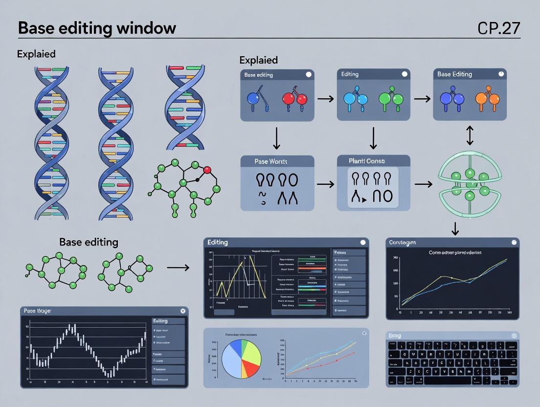
What is the Base Editing Window? Core Principles and Biochemical Foundations
Within the context of a broader thesis on "Base editing window explained," this technical guide elucidates the principle of the editing window as a dynamic profile of enzymatic activity across a stretch of target DNA, rather than a binary on-target site. This concept is critical for the precise application of base editors (BEs) in therapeutic development and functional genomics.
Base editors are fusion proteins combining a catalytically impaired Cas nuclease with a nucleobase deaminase enzyme. Their activity is not confined to a single nucleotide but spans a region of single-stranded DNA within the R-loop formed by Cas9 binding. This region of potential deamination is termed the "editing window." Its definition is probabilistic, determined by the accessibility of substrate nucleotides to the deaminase's active site and the kinetics of the entire complex.
Quantitative Profiling of Editing Windows
The editing window is experimentally defined by high-throughput sequencing of edited populations, quantifying the percentage of reads with a specific base conversion at each position within the protospacer. Data is typically presented as an activity profile.
Table 1: Representative Editing Window Characteristics for Common Base Editors
| Base Editor Type | Deaminase Domain | Typical Window (positions from PAM, NGG) | Primary Conversion | Average Peak Efficiency (%)* | Key Determinant of Window Width |
|---|---|---|---|---|---|
| BE3 / BE4max | rAPOBEC1 | ~Positions 4-10 (C•G to T•A) | C to T | 30-70 | Linker length & flexibility |
| ABE7.10 / ABE8e | TadA-7.10/TadA-8e | ~Positions 4-9 (A•T to G•C) | A to G | 40-80 | Deaminase processivity |
| CRISPR-X / SECURE | hA3A / hA3B | ~Positions 1-17 (broad) | C to T | 5-50 | Deaminase sequence preference |
| Target-AID | PmCDA1 | ~Positions 1-7 (C•G to T•A) | C to T | 10-40 | Deaminase processivity |
Note: Efficiency is highly target-sequence dependent. Values represent common ranges from model genomic loci.
Core Experimental Protocol for Defining an Editing Window
This methodology outlines the standard workflow for empirically determining a base editor's editing window at a novel genomic locus.
Protocol: Amplicon Sequencing-Based Editing Window Analysis
- Design & Cloning: Design a single guide RNA (sgRNA) targeting the locus of interest. Clone the sgRNA sequence into an appropriate expression plasmid (e.g., pX330 derivative for SpCas9).
- Cell Transfection: Co-transfect HEK293T cells (or relevant cell line) with the sgRNA plasmid and the base editor expression plasmid using a standard transfection reagent (e.g., Lipofectamine 3000). Include a negative control (sgRNA only).
- Genomic DNA Harvest: 72 hours post-transfection, harvest cells and extract genomic DNA using a silica-column based kit.
- PCR Amplification: Design primers flanking the target site (~250-300 bp amplicon). Perform PCR using a high-fidelity polymerase.
- Amplicon Library Prep & Sequencing: Purify PCR products, barcode samples using a dual-indexing strategy (e.g., Nextera XT), and pool for sequencing on an Illumina MiSeq or NextSeq platform (2x150 bp or 2x250 bp).
- Data Analysis:
- Alignment: Demultiplex reads and align to the reference genome using tools like BWA or CRISPResso2.
- Variant Calling: Use specialized tools (e.g., CRISPResso2, BE-Analyzer) to quantify the percentage of sequencing reads containing C>T (or A>G) conversions at every position within the target amplicon.
- Visualization: Plot conversion frequency (%) against nucleotide position relative to the PAM to generate the activity profile.
Visualizing the Concept and Workflow
The Scientist's Toolkit: Key Research Reagent Solutions
Table 2: Essential Reagents for Editing Window Analysis
| Reagent / Material | Function / Role in Experiment | Example Product / Note |
|---|---|---|
| Base Editor Expression Plasmid | Delivers the BE (e.g., BE4max, ABE8e) to cells. | Addgene #112093 (BE4max), #138489 (ABE8e). |
| sgRNA Cloning Backbone | Plasmid for expressing the target-specific guide RNA. | Addgene #62988 (pX330-U6-Chimeric_BB-CBh-hSpCas9). |
| High-Efficiency Transfection Reagent | Enables delivery of plasmids into mammalian cells. | Lipofectamine 3000, FuGENE HD. |
| Genomic DNA Extraction Kit | Purifies high-quality gDNA for PCR amplification. | DNeasy Blood & Tissue Kit (Qiagen), Quick-DNA Miniprep Kit (Zymo). |
| High-Fidelity PCR Polymerase | Amplifies target locus with minimal error for NGS. | Q5 Hot-Start (NEB), KAPA HiFi HotStart. |
| NGS Library Prep Kit | Prepares barcoded amplicon libraries for sequencing. | Illumina Nextera XT, Swift Biosciences Accel-NGS 2S. |
| Analysis Software | Quantifies base conversion frequencies from NGS data. | CRISPResso2, BE-Analyzer, or custom Python/R scripts. |
Implications for Drug Development and Research
Understanding the editing window is non-negotiable for therapeutic applications. A broad window increases the risk of bystander edits (unwanted conversions within the window), potentially creating pathogenic mutants. Conversely, a narrow, predictable window is ideal for correcting point mutations. Current research focuses on engineering BEs with narrowed or tunable windows through directed evolution of the deaminase domain, rational linker design, and the use of Cas variants with altered conformational dynamics.
Within the broader thesis of base editing window explained research, a central mechanistic question persists: what structural and geometric factors constrain the catalytic efficiency and sequence specificity of deaminase enzymes bound to RNA or DNA substrates? This whitepaper provides an in-depth technical guide to the structural biology insights that reveal how the three-dimensional architecture of deaminase-substrate complexes imposes stringent constraints, thereby defining the editable "window" in base editing technologies. Understanding these geometric constraints is paramount for researchers and drug development professionals aiming to engineer next-generation editors with enhanced precision and expanded therapeutic utility.
Structural Architecture of Deaminase-Substrate Complexes
Deaminases used in base editing, such as APOBEC and AID families for cytidine deamination or TadA variants for adenosine deamination, share a common core fold but exhibit distinct modes of nucleic acid recognition. The geometry of the complex is governed by:
- Active Site Pocket Dimensions: The physical volume and electrostatic landscape of the catalytic pocket dictate which nucleoside can be positioned for deamination. Mismatches in size or charge preclude catalysis.
- Substrate Strand Conformation: The nucleic acid often undergoes severe kinking or distortion, typically involving a flipped-out target nucleoside that rotates into the active site. The energy required for this extrusion is sequence-context-dependent.
- Accessory Domain Interactions: Cas-derived proteins in CRISPR-base editor fusions, or RNA-binding domains in standalone deaminases, position the catalytic domain relative to the substrate strand, setting the initial geometric parameters.
Table 1: Quantitative Geometric Parameters of Characterized Deaminase Complexes
| Deaminase Family | PDB Code (Example) | Target Base | Base Flip Angle (°) | Catalytic Pocket Volume (ų) | Key Constraining Residue(s) | Measured Editing Window (nt) |
|---|---|---|---|---|---|---|
| APOBEC3A | 5SWW | Cytidine | ~180 | ~540 | W104, P210 | ~5 (ssDNA) |
| TadA-8e (ABE8e) | 7NJ4 | Adenosine | ~165 | ~610 | D108, Y147 | ~4-5 (within R-loop) |
| AID | 5JJJ | Cytidine | ~170 | ~520 | R112, P151 | N/A |
How Geometry Imposes Catalytic Constraints
The precise alignment of the target base with the catalytic zinc ion and water molecule is non-negotiable. Geometric constraints arise from:
- Steric Exclusion: Side chains lining the pocket (e.g., tryptophan, tyrosine) create a "molecular ruler" that excludes bases with larger functional groups or incorrect tautomeric forms.
- Backbone Torsion Stress: The phosphodiester backbone of the substrate must adopt high-energy conformations to achieve base extrusion. Neighboring sequences that resist this distortion (e.g., high GC content, secondary structure) reduce editing efficiency, creating a de facto window constraint.
- Spacing Limitations in Fused Editors: In CRISPR-Base Editors, the rigid linker length between Cas9 and deaminase, combined with the width of the Cas9-induced R-loop, defines a strict spatial zone (typically protospacer positions 4-8) where the deaminase active site can access the DNA strand.
Experimental Protocols for Elucidating Geometric Constraints
X-ray Crystallography of Deaminase-Substrate Complexes
Protocol:
- Protein Expression & Purification: Express recombinant deaminase (with solubility tags) in E. coli or insect cells. Purify via affinity (Ni-NTA/Co²⁺ for His-tag), ion-exchange, and size-exclusion chromatography.
- Oligonucleotide Preparation: Synthesize and HPLC-purify short DNA or RNA substrates containing a target base. Anneal to complementary strands if needed.
- Complex Formation & Crystallization: Incubate protein and oligonucleotide at a 1:1.2 molar ratio. Screen for crystals using commercial sparse-matrix screens (e.g., Hampton Research) in sitting-drop vapor diffusion plates at 4°C and 20°C.
- Data Collection & Refinement: Flash-freeze crystals in liquid N₂ with cryoprotectant. Collect diffraction data at a synchrotron beamline. Solve structure via molecular replacement using a related deaminase structure. Iteratively refine model (e.g., with PHENIX) and validate.
Cryo-EM for Large Fused Editor Complexes
Protocol:
- Sample Preparation: Assemble full-length base editor (e.g., BE4max) with sgRNA and target DNA duplex in vitro. Apply 3-4 µL of sample to glow-discharged cryo-EM grids, blot, and plunge-freeze in liquid ethane.
- Data Acquisition: Collect multi-frame movies on a 300 keV cryo-TEM with a K3 direct electron detector. Target a defocus range of -0.8 to -2.5 µm. Use beam-image shift to collect multiple holes per stage movement.
- Image Processing: Motion-correct and dose-weight frames. Perform template-based particle picking, 2D classification, ab-initio reconstruction, and heterogeneous refinement in cryoSPARC or RELION. Sharpen the final map and build an atomic model using Coot and real-space refine.
Molecular Dynamics (MD) Simulations
Protocol:
- System Setup: Start from a crystallographic or cryo-EM model. Solvate the complex in a TIP3P water box with 150 mM NaCl. Neutralize system charge.
- Energy Minimization & Equilibration: Minimize energy for 10,000 steps using the CHARMM36 force field. Gradually heat the system from 0 to 310 K over 100 ps in an NVT ensemble, then equilibrate at 1 atm for 1 ns in an NPT ensemble.
- Production Run & Analysis: Run unrestrained MD for 100-500 ns. Analyze trajectories for root-mean-square deviation (RMSD), fluctuation (RMSF), hydrogen bonding, and base-flipping dynamics using VMD/NAMD or GROMACS suites.
Visualization of Structural Constraints and Editing Workflow
Base Editor Geometric Constraint Workflow
Structural Constraint Logic Map
The Scientist's Toolkit: Research Reagent Solutions
Table 2: Essential Reagents for Structural Studies of Deaminase Constraints
| Reagent / Material | Function in Research | Example Product / Vendor |
|---|---|---|
| Recombinant Deaminase Protein | High-purity, active enzyme for crystallography, biochemistry, and complex assembly. | Custom expression in E. coli BL21(DE3), purification via HisTrap HP (Cytiva). |
| Chemically Modified Oligonucleotides | Substrates with non-hydrolyzable analogs (e.g., 2'-fluoro) to trap intermediates for structural studies. | Custom synthesis from IDT or Thermo Fisher. |
| Crystallization Screening Kits | Identify initial conditions for growing protein-nucleic acid co-crystals. | JC SG Core Suites I-IV (Qiagen), Morpheus (Molecular Dimensions). |
| Cryo-EM Grids | Support film for vitrifying large macromolecular complexes for EM. | Quantifoil R1.2/1.3 Au 300 mesh (Electron Microscopy Sciences). |
| MD Simulation Software | Perform all-atom simulations to analyze dynamics and energy landscapes. | GROMACS (Open Source), AMBER (Commercial). |
| Surface Plasmon Resonance (SPR) Chip | Measure binding kinetics and affinity between deaminase and substrate variants. | Series S Sensor Chip NTA (Cytiva). |
Within the broader thesis of base editing window research, the spatial and functional characteristics of the editing window are paramount. This technical guide elucidates the core engineering parameters—single-guide RNA (sgRNA) length, linker design, and deaminase variant selection—that collaboratively define the width, position, and efficiency of the base editing activity window. Understanding these factors is critical for researchers, scientists, and drug development professionals to design precise and predictable base editing systems.
Base editors (BEs) are engineered fusion proteins that enable the direct, irreversible conversion of one DNA base pair to another without generating double-strand breaks. The editing "window" refers to the span of DNA nucleotides within the protospacer where deamination activity occurs with significant efficiency. The window's size (width) and positional offset from the protospacer-adjacent motif (PAM) are not fixed but are tunable variables directly influenced by protein and RNA engineering.
sgRNA Length: Defining the Scaffold and Spacing
The sgRNA length, particularly the length of the spacer sequence, is a primary determinant of the spatial relationship between the deaminase active site and the target nucleotide.
- Standard Length (20 nt): Conventional sgRNAs utilize a 20-nucleotide spacer. This places the deaminase domain's window typically within a zone spanning approximately positions 4-8 (for cytosine base editors, CBEs) or 3-10 (for adenine base editors, ABEs) from the PAM-distal end of the protospacer.
- Truncated or Extended Spacers: Altering spacer length shifts the position of the entire ribonucleoprotein complex relative to the PAM, thereby translating the editing window. Shorter spacers (e.g., 16-18 nt) can shift the window closer to the PAM, while longer spacers can shift it further away. However, extreme deviations can reduce binding affinity and overall editing efficiency.
Table 1: Impact of sgRNA Spacer Length on Editing Window Position
| Spacer Length (nt) | Effect on RNP Architecture | Typical Window Shift (Relative to 20-nt Standard) | Primary Application |
|---|---|---|---|
| 16-17 | Compacts complex, brings deaminase closer to PAM | Window shifts ~2-4 bases toward PAM | Editing sites very close to PAM. |
| 18-19 | Moderate compaction. | Window shifts ~1-2 bases toward PAM. | Fine-tuning for optimal activity. |
| 20 | Standard architecture. | Reference position (e.g., CBE window ~ed4-8). | General-purpose editing. |
| 21-23 | Extends reach of deaminase away from PAM. | Window shifts ~1-3 bases away from PAM. | Accessing distal sites within a protospacer. |
Linker Design: The Mechanical Coupling
The linker tethering the deaminase domain to the Cas9 nickase (nCas9) or dead Cas9 (dCas9) is a critical mechanical component. Its length, flexibility, and composition govern the permissible "reach" and rotational freedom of the deaminase, directly impacting window width and profile.
- Length: Shorter linkers restrict the deaminase's rotational and translational motion, often narrowing the editing window. Longer linkers increase flexibility and can widen the window but may also reduce overall efficiency due to entropy.
- Composition & Rigidity: Flexible linkers (e.g., (GGGS)n repeats) allow a broad exploration space, potentially widening the window. More rigid or structured linkers can constrain the deaminase to a more precise spatial envelope, narrowing the window.
Table 2: Linker Properties and Their Impact on Editing Window
| Linker Property | Example Sequences/Structures | Impact on Window | Rationale |
|---|---|---|---|
| Short & Flexible | (GGGS)_1-2 | Narrower, more defined window. | Restricted spatial sampling of deaminase. |
| Long & Flexible | (GGGS)3-5, (X)n linkers | Potentially wider, diffuse window. | Increased range of motion for deaminase domain. |
| Rigid/Structured | α-helical linkers, protein domains | Alters window position/profile; can narrow. | Constrains deaminase orientation precisely. |
| Optimized Hybrid | e.g., "XTEN" linkers, designed sequences | Tunable for balance of width/efficiency. | Engineered for specific biophysical properties. |
Deaminase Variant: The Catalytic Engine
The choice of deaminase and its engineered variants is the most potent factor for modulating window characteristics. Different deaminases have intrinsic structural preferences for ssDNA substrates, and directed evolution has created variants with altered window properties.
- rAPOBEC1 (CBE): The canonical deaminase for CBEs. Early BE4 variants exhibit a window of ~positions 4-8. Evolved variants like BE4max improve efficiency without drastically altering the window.
- AID/APOBEC Family Variants: Deaminases like AID, A3A, and A3B have different sequence context preferences and can exhibit wider or narrower windows in their base editor fusions.
- TadA (ABE): The laboratory-evolved E. coli tRNA deaminase TadA-8e is the core of modern ABEs. The window for ABE8e variants is typically broader (~positions 3-10) compared to earlier ABE7.10 versions. Further engineering (e.g., ABE8e-SpRY) can expand targetable sites when combined with PAM-relaxed Cas variants.
Table 3: Deaminase Variants and Associated Window Profiles
| Base Editor | Core Deaminase Variant | Typical Window (Positions from PAM-distal end, 20-nt spacer) | Key Characteristics |
|---|---|---|---|
| BE4, BE4max | rAPOBEC1 (evolved) | 4-8 (C4-C8) | Standard high-efficiency CBE. |
| A3A-BE | human APOBEC3A | 2-6 (C2-C6) | Narrower window, high on-target efficiency. |
| Target-AID | pmCDA1 (AID-like) | 1-7 (C1-C7) | Broader window, can have higher off-target RNA editing. |
| ABE7.10 | TadA-7.10/TadA-8e heterodimer | 4-7 (A4-A7) | Original ABE, relatively narrow window. |
| ABE8e | TadA-8e homodimer | 3-10 (A3-A10) | Broadened window, significantly higher activity. |
Integrated Experimental Protocol: Determining Window Characteristics
Objective: To empirically define the editing window of a novel base editor construct (e.g., combining a new linker with a deaminase variant). Workflow:
- Target Design: Select a genomic locus with a neutral sequence context. Design a panel of 8-10 sgRNAs targeting the same strand, each shifting the target protospacer by 1 base relative to a fixed PAM. Include sgRNAs with varying spacer lengths (17, 18, 20, 22 nt) for a subset.
- Transfection: Co-transfect HEK293T cells (or relevant cell line) with a constant amount of the base editor plasmid and each individual sgRNA expression plasmid (or a pooled library for NGS-based assays), in triplicate.
- Harvest & Amplification: Harvest genomic DNA 72-96 hours post-transfection. Perform PCR amplification of the target locus using barcoded primers to allow for multiplexed sequencing.
- Sequencing & Analysis: Perform high-depth amplicon sequencing (NGS). Align reads to the reference sequence. Calculate the percentage of sequencing reads exhibiting each possible base conversion (C-to-T or A-to-G) at every position within the protospacer and surrounding region.
- Window Determination: Plot editing efficiency (%) versus genomic position. The editing window is defined as the contiguous set of positions where efficiency exceeds a predetermined threshold (e.g., 5% or 10% of total reads). The width is the number of bases in this set, and the position is defined relative to the PAM.
Diagram Title: Base Editor Window Characterization Workflow
The Scientist's Toolkit: Research Reagent Solutions
Table 4: Essential Reagents for Base Editing Window Research
| Reagent/Kit | Function/Application | Key Considerations |
|---|---|---|
| Base Editor Expression Plasmids | Delivery of BE machinery. Common backbones: pCMV-BE4max, pCMV-ABE8e. | Ensure promoter is active in your cell type (CMV, EF1α, CAG). |
| sgRNA Cloning Kit | Rapid assembly of sgRNA expression constructs (e.g., into U6 promoter vectors). | Golden Gate assembly (BsaI) or annealed-oligo cloning are standard. |
| High-Efficiency Transfection Reagent | Delivery of plasmids to mammalian cells (e.g., Lipofectamine 3000, PEI). | Optimize for your cell line; primary cells often require specialized methods. |
| NGS Amplicon-EZ Service/Library Prep Kit | Preparation of PCR amplicons for Illumina sequencing. | Services from Azenta/Genewiz or kits from Illumina/NEB streamline the process. |
| CRISPR Analysis Software (e.g., CRISPResso2, BE-Analyzer) | Quantification of base editing efficiency from NGS data. | Critical for accurate, batch-processed analysis of window profiles. |
| Surveyor/T7 Endonuclease I Kits | Lower-throughput alternative for detecting editing-induced mismatches. | Less quantitative and not base-specific compared to NGS. |
| Sanger Sequencing & EditR/TIDE Analysis | Rapid, low-cost assessment of editing at single sites. | Useful for initial validation but lacks the resolution for full window profiling. |
Diagram Title: Core Factors Converge to Define the Editing Window
The base editing window is a malleable property, not a fixed constraint. By systematically engineering the tripartite system of sgRNA length (for positioning), linker design (for mechanical leverage), and deaminase variant (for catalytic specificity and processivity), researchers can tailor the window's size and location to suit specific therapeutic or research applications. This rational design approach, framed within the ongoing thesis of base editing optimization, is fundamental to advancing the precision and utility of base editing technologies in biomedicine.
This whitepaper explores a critical advancement in base editing research: the distinction between the canonical editing window, a predictable region derived from structural and biochemical models of the editor complex, and the real-world editing window, which is empirically measured and influenced by genomic context, chromatin state, and cellular delivery. Understanding and reconciling this dichotomy is essential for optimizing the efficacy and safety of base editors in therapeutic and research applications. This document is framed within the broader thesis that precise definition of the "base editing window" is not a fixed property of the editor alone, but a dynamic outcome of its interaction with the genome.
Defining the Editing Windows
Canonical Editing Window: This is the theoretical, sequence-agnostic region within the single-stranded DNA bubble (R-loop) formed during Cas9 binding where the deaminase domain has steric and catalytic access to the target nucleobase. For common cytosine base editors (CBEs), this is typically positions 4-8 (counting the PAM as positions 21-23). For some adenine base editors (ABEs), it is positions 4-7. This window is predicted from crystallography and in vitro biochemical assays.
Real-World Editing Window: This is the experimentally observed distribution of base conversions across the target site in living cells or complex in vitro systems. It deviates from the canonical window due to factors such as:
- Sequence Context: Local DNA sequence affects deaminase activity and processivity.
- Chromatin Accessibility: Nucleosome occupancy and histone modifications restrict physical access.
- Cellular Delivery & Expression: The method (e.g., viral transduction, electroporation) and duration of editor expression influence outcomes.
- DNA Repair Dynamics: Cellular repair pathways can mask or correct edits non-uniformly.
Quantitative Data Comparison
The following tables summarize key quantitative differences between canonical predictions and real-world measurements for common base editors.
Table 1: Theoretical vs. Observed Editing Windows for Common Base Editors
| Base Editor | Canonical Window (Positions from PAM) | Typical Real-World Window (Observed Range) | Average Peak Efficiency Discrepancy |
|---|---|---|---|
| BE4 (CBE) | 4-8 | 3-10 | Canonical predicts ~80% at pos5; Real-world often shows 40-60% due to context. |
| ABE8e (ABE) | 4-7 | 4-9 | Broader activity, with significant editing at position 9 not predicted by canonical model. |
| CRISPR-Cas12a CBE | 8-13 (from PAM) | 7-16 | Greater spread, with strong influence from sequence-specific deaminase preference. |
Table 2: Factors Causing Real-World vs. Canonical Discrepancies & Measured Impact
| Influencing Factor | Experimental Impact on Window Width/Position | Typical Measurement Method |
|---|---|---|
| GC Content | High GC >5% narrowing of window, shift in peak. | Deep sequencing of synthetic target libraries. |
| Chromatin State (Closed vs Open) | Closed chromatin can reduce efficiency >90%, distorting window shape. | ATAC-seq correlation with editing efficiency. |
| sgRNA Spacer Length | 20-nt vs 18-nt spacer can shift window by 1-2 nucleotides. | Parallel screening with truncated spacers. |
| Delivery Modality (LNP vs AAV) | AAV persistence leads to broader, less precise windows over time. | Longitudinal tracking via NGS. |
Experimental Protocols for Measurement
Protocol 1: High-Throughput Determination of Real-World Editing Windows
Objective: Empirically map the real-world editing window for a novel base editor across diverse genomic contexts.
Methodology:
- Library Design: Synthesize an oligo pool containing the target editor scaffold flanking a randomized 20-nt window (covering positions 1-20 relative to PAM). Clone this library into a lentiviral backbone with a barcode for sequencing.
- Cell Delivery: Transduce the library into HEK293T cells at low MOI. Transfect with plasmid expressing the base editor and a constant sgRNA targeting the scaffold.
- Harvest & Sequencing: Harvest genomic DNA 72h post-transfection. Amplify the target region with primers containing Illumina adapters and unique molecular identifiers (UMIs).
- Data Analysis: Align sequences to reference. Quantify base conversion frequencies at each position for each sequence context. Plot editing efficiency as a function of position to define the real-world window.
Protocol 2: Chromatin Accessibility Correlation Assay
Objective: Measure the impact of native chromatin state on the observed editing window.
Methodology:
- Target Selection: Choose 50 genomic loci with known, varying ATAC-seq signal (open, intermediate, closed).
- Parallel Editing: Deliver the same base editor (e.g., ABE8e) and locus-specific sgRNAs to cells via nucleofection.
- Dual Harvest: Split cells 72h post-editing. One aliquot for gDNA extraction (editing analysis by amplicon sequencing). The other for ATAC-seq to confirm accessibility state.
- Correlative Analysis: Plot per-locus editing efficiency (and window shape) against its normalized ATAC-seq read count. Fit a regression model to quantify the accessibility effect.
Visualizations
Diagram Title: Conceptual Relationship Between Editing Window Definitions
Diagram Title: High-Throughput Real-World Window Mapping Workflow
The Scientist's Toolkit: Research Reagent Solutions
Table 3: Essential Reagents for Editing Window Studies
| Reagent / Material | Function in Protocol | Key Consideration |
|---|---|---|
| Synthetic Oligo Pool Library | Provides diverse sequence context to test editor activity. | Ensure high complexity and balanced nucleotide representation. |
| Lentiviral Packaging System | For stable genomic integration of target library. | Use 3rd generation system for biosafety; titer carefully. |
| High-Fidelity DNA Polymerase | For error-free amplification of pre- and post-editing sequences. | Critical for accurate variant frequency quantification. |
| Unique Molecular Identifiers (UMIs) | Short random nucleotide tags to correct for PCR amplification bias. | Essential for accurate quantitative NGS. |
| Validated Base Editor Expression Plasmid | Consistent source of editor protein. | Use a strong, constitutive promoter (e.g., CAG, EF1α). |
| ATAC-seq Kit | To measure chromatin accessibility in parallel with editing. | Use fresh cells or cryopreserved nuclei for best results. |
| Single-Guide RNA (sgRNA) | Directs base editor to target locus. | Chemical modifications can enhance stability and efficiency. |
| Next-Generation Sequencing Platform | For deep sequencing of target amplicons. | Aim for >10,000x coverage per sample for statistical power. |
Within the context of base editing, the "editing window" refers to the specific span of DNA nucleotides within the protospacer where the deaminase enzyme can catalyze a base conversion. This window is primarily constrained by the steric limitations of the Cas9-deaminase fusion protein and the accessibility of the single-stranded DNA within the R-loop structure. The precise boundaries and efficiency profile of this window are not uniform; they are dictated by the specific base editor architecture (e.g., BE4, ABE8e), the guide RNA (gRNA) sequence, and the local chromatin context. Understanding and controlling this window is the central thesis of modern base editing optimization, as it directly dictates the balance between achieving the desired on-target edit and minimizing unwanted, promiscuous deamination.
Quantitative Analysis of Window Characteristics
The following tables summarize key quantitative data from recent studies characterizing editing windows for prevalent base editor systems.
Table 1: Characteristic Editing Windows of Common Base Editors
| Base Editor System | Deaminase Type | Primary Conversion | Typical Window Position (Protospacer, 5'→3') | Peak Efficiency Within Window | Key Reference (Example) |
|---|---|---|---|---|---|
| BE4max | rAPOBEC1 | C•G to T•A | Positions 4-8 (≈ spacer nucleotides 4-8) | Positions 5-7 | Komor et al., 2017; Rees et al., 2019 |
| ABE8e | TadA-8e | A•T to G•C | Positions 4-8 (≈ spacer nucleotides 4-8) | Positions 4-7 | Richter et al., 2020 |
| Target-AID | PmCDA1 | C•G to T•A | Positions 1-6 (≈ spacer nucleotides 1-6) | Positions 2-5 | Nishida et al., 2016 |
| CRISPR-Cas12a BE | rAPOBEC1 | C•G to T•A | Positions 6-13 (post-PAM) | Positions 8-10 | Li et al., 2018 |
| SECURE-BE3 (mutant) | rAPOBEC1* | C•G to T•A | Positions 4-8 (with reduced off-target) | Positions 5-7 | Yu et al., 2020 |
Table 2: Correlation Between Window Position and Byproduct Frequencies
| Editing Position (from PAM) | Relative Deamination Efficiency | Indel Frequency (%) | Typical Undesired Byproducts (CBE Example) |
|---|---|---|---|
| 3-4 | Low to Moderate | <0.5% | Low, but possible non-C-to-T edits |
| 5-7 (Peak) | Very High | 0.5 - 2.0% | Higher risk of C-to-G, C-to-A ("bystanders") |
| 8-10 | Moderate | 1.0 - 3.0% | Increased stochastic indels |
| >12 | Very Low | Variable | Primarily background noise |
Experimental Protocols for Window Characterization
Protocol 1: High-Throughput Sequencing Analysis of Editing Window Profile
Objective: To quantitatively determine the efficiency and product distribution at each nucleotide position within the potential editing window for a given base editor and gRNA.
Methodology:
- Design & Transfection: Design a gRNA targeting a genomically integrated or synthetic locus containing multiple target bases within the protospacer. Co-transfect HEK293T cells (or relevant cell line) with plasmids encoding the base editor and the gRNA using a standard method (e.g., PEI, Lipofectamine 3000).
- Harvest & Amplification: Harvest genomic DNA 72 hours post-transfection. Perform PCR amplification of the target locus using high-fidelity polymerase, with primers containing Illumina adapter overhangs.
- Library Prep & Sequencing: Index the amplicons via a second limited-cycle PCR and purify. Pool libraries for deep sequencing on an Illumina MiSeq or NextSeq platform (aim for >50,000x read depth per sample).
- Data Analysis: Process FASTQ files using a pipeline (e.g., CRISPResso2, BE-Analyzer). Align reads to the reference sequence. Calculate the percentage of reads with a C-to-T (for CBE) or A-to-G (for ABE) conversion at each position within the protospacer. Also quantify frequencies of indels and other base substitutions (byproducts).
Protocol 2: In Vitro Deamination Assay for Window Definition
Objective: To delineate the intrinsic biochemical window of a base editor independent of cellular processes like DNA repair.
Methodology:
- Substrate Preparation: Generate a double-stranded DNA substrate containing the target sequence with a 5' fluorescent label (e.g., FAM) on one strand. Alternatively, use a synthetic, partially duplexed DNA oligonucleotide mimicking the R-loop structure.
- Protein Purification: Purify the base editor protein (e.g., BE4) and Cas9 nuclease (as control) via affinity chromatography (e.g., His-tag, MBP-tag).
- Reaction Setup: Incubate the DNA substrate (50 nM) with the purified base editor (100 nM) and a matched gRNA (120 nM) in reaction buffer (e.g., 20 mM HEPES pH 7.5, 150 mM KCl, 5 mM MgCl2, 1 mM DTT) at 37°C for 60 minutes.
- Analysis: Terminate the reaction and denature DNA. Analyze products by capillary electrophoresis (if fluorescently labeled) or next-generation sequencing of the reaction products. The readout shows deamination events directly catalyzed within the enzyme's accessible window.
Visualizing Concepts and Workflows
Title: Base Editing Workflow & Risk Pathways
Title: CBE vs ABE Editing Window Efficiency Profiles
The Scientist's Toolkit: Research Reagent Solutions
| Item | Function / Relevance to Window Studies |
|---|---|
| BE4max Plasmid (Addgene #112093) | A high-efficiency CBE variant. Standard tool for establishing baseline CBE window characteristics (positions 4-8). |
| ABE8e Plasmid (Addgene #138489) | A high-activity ABE variant. Used to define the optimized A-to-G editing window and compare to CBEs. |
| CRISPResso2 Software | Computational tool for deep sequencing analysis. Crucial for quantifying editing percentages at each nucleotide position. |
| Synthetic gRNA (chemically modified) | Enhances stability and editing efficiency. Using a consistent, high-quality gRNA is vital for reproducible window profiling. |
| HEK293T Cell Line | A standard, highly transfectable mammalian cell line used for initial characterization of editor performance and window. |
| KAPA HiFi HotStart PCR Kit | Provides high-fidelity amplification of target loci for NGS library preparation, minimizing PCR-induced errors. |
| Illumina DNA Prep Kit | Streamlined library preparation for amplicon sequencing, enabling high-throughput screening of editing outcomes. |
| Recombinant BE Protein (NEB #E3323S) | Purified base editor for in vitro assays. Allows precise biochemical definition of the deamination window without cellular confounders. |
| Sanger Sequencing (ACGT Corp.) | For rapid, initial validation of editing success and rough estimation of primary editing site efficiency. |
| Guide Design Tool (Benchling) | In-silico design and specificity checking of gRNAs, helping to avoid promiscuous windows in homologous genomic regions. |
Characterizing and Harnessing the Editing Window: Experimental Strategies and Design Rules
Within the broader thesis of "Base editing window explained research," profiling the activity landscape of base editors (BEs) across a genomic target is paramount. The "editing window"—the region of nucleotides within a protospacer where efficient base conversion occurs—is a critical determinant of editing precision, specificity, and therapeutic viability. This technical guide details standard assays for comprehensive window profiling, leveraging deep sequencing and robust Next-Generation Sequencing (NGS) analysis pipelines to quantitatively map editor performance.
Core Deep Sequencing Approaches for Window Profiling
Accurate window profiling requires sequencing assays that capture both the identity and frequency of editing events at single-nucleotide resolution across entire amplicons.
Targeted Amplicon Sequencing (Amp-Seq)
This is the gold-standard method for quantifying editing outcomes at defined genomic loci.
- Principle: Genomic DNA encompassing the target site is PCR-amplified, barcoded with unique dual indices (UDIs), and sequenced at high depth (>10,000x coverage).
- Key Advantage: Provides quantitative data on base substitution frequencies, insertion/deletion (indel) rates, and byproduct formations (e.g., undesired transversions, bystander edits) for every position in the amplicon.
High-Throughput Window Profiling with Saturated Targeting
To systematically define editing windows, researchers employ libraries of single-guide RNAs (sgRNAs) targeting a locus with tiling spacers or saturated mutagenesis of a single spacer.
- Principle: A plasmid library encoding thousands of sgRNA variants targeting a region of interest is co-delivered with the base editor. NGS of both the sgRNA library (to assess representation) and the corresponding genomic targets (to assess outcomes) allows for the parallel measurement of editing efficiency across numerous sequence contexts.
NGS Analysis Pipelines: From Raw Data to Window Metrics
A standardized bioinformatics workflow is essential for transforming raw sequencing reads into interpretable window profiling data.
Diagram 1: NGS Analysis Pipeline for Window Profiling
Diagram Title: Workflow for Base Editing NGS Data Analysis
Detailed Protocol: Amp-Seq Data Analysis with CRISPResso2
Input: Paired-end FASTQ files from sequencing of the target amplicon.
- Preprocessing & Alignment: Use Cutadapt to trim primer sequences. Align reads to a reference amplicon sequence using Bowtie2.
- Quantification: Utilize CRISPResso2 in "AMP-Seq" or "Base Editing" mode.
- Command Example:
- Command Example:
- Output: CRISPResso2 generates a table (
Quantification_of_editing_frequency.txt) reporting the percentage of reads with each nucleotide at every position. This is the primary data for window profiling.
Key Metrics for Window Characterization
From the quantification table, calculate:
- Primary Editing Efficiency: % of reads with the intended base change at the target position.
- Product Purity: (% intended product) / (% intended product + % byproducts) at the target position.
- Bystander Editing Efficiency: % of reads with the intended base change at non-target positions within the window.
- Editing Window Width: The contiguous set of positions where editing efficiency exceeds a predefined threshold (e.g., 5% or 10% of the peak efficiency).
Table 1: Representative Window Profiling Data for Common Base Editors (Hypothetical Data)
| Base Editor | Target Base Change | Peak Efficiency (%) | Product Purity (%) | Editing Window (Positions)* | Avg. Bystander Efficiency within Window (%) |
|---|---|---|---|---|---|
| BE4 | C•G to T•A | 65 | 88 | 4-8 | 12 |
| ABE8e | A•T to G•C | 80 | 95 | 4-7 | 5 |
| CGBE1 | C•G to G•C | 45 | 75 | 3-9 | 18 |
*Positions are relative to the protospacer adjacent motif (PAM), typically numbered as PAM-distal (1) to PAM-proximal (~20).
Table 2: Comparative NGS Analysis Tools for Window Profiling
| Tool Name | Primary Function | Key Inputs | Outputs for Window Profiling | Best For |
|---|---|---|---|---|
| CRISPResso2 | Quantification of editing outcomes | FASTQ, Amplicon Seq, Expected Edit | Efficiency by position, allele tables, plots | Standard Amp-Seq, detailed bystander analysis |
| BE-Analyzer | Specialized for base editor analysis | FASTQ, Reference, BED file | Normalized editing rates, window graphs | High-throughput tiling sgRNA screens |
| CRISPResso2WGS | Genome-wide specificity analysis | Whole Genome Sequencing (WGS) data | Off-target candidate sites, potential bystanders | Genome-wide window profiling for off-targets |
The Scientist's Toolkit: Key Research Reagent Solutions
Table 3: Essential Materials for Base Editing Window Profiling Experiments
| Item | Function & Explanation |
|---|---|
| Validated Base Editor Plasmid (e.g., pCMV_BE4) | Expression construct for the base editor protein. Ensures consistent editor delivery and activity. |
| sgRNA Cloning Backbone (e.g., pU6-sgRNA) | Vector for expressing the target-specific single-guide RNA. |
| NGS-Amplicon PCR Primers with Overhang Adapters | Primers containing Illumina sequencing adapter overhangs for direct library preparation from genomic DNA. |
| High-Fidelity DNA Polymerase (e.g., Q5, KAPA HiFi) | For error-free amplification of the target locus prior to sequencing. Critical for accurate variant calling. |
| Dual-Indexed UMI Adapter Kit (e.g., Illumina TruSeq) | Allows multiplexing of samples and incorporation of Unique Molecular Identifiers (UMIs) for accurate deduplication. |
| Genomic DNA Extraction Kit (Cell Culture/ Tissue) | To obtain high-quality, RNase-free genomic DNA from edited samples. |
| CRISPResso2 Software Package | Core bioinformatics tool for quantifying base editing outcomes from NGS data. |
| Reference Genome FASTA File | Species-specific reference genome sequence for accurate read alignment. |
| Validated Positive Control sgRNA | An sgRNA with known high editing efficiency to control for editor performance in each experiment. |
Advanced Pathway: Integrating Window Data with Functional Outcomes
For therapeutic development, editing window data must be integrated with functional annotations.
Diagram 2: From Window Profile to Therapeutic Design
Diagram Title: Integrating Window Profiling into Therapeutic Design
Protocol: In Silico sgRNA Selection using Window Data:
- Overlay: Map the high-efficiency editing window (e.g., positions 4-8 for BE4) onto the target genomic region of interest.
- Annotate: Annotate each nucleotide in the window using databases (e.g., UCSC Genome Browser) to identify:
- Target pathogenic nucleotide (e.g., disease-causing C•G).
- Critical bystander nucleotides (e.g., a C in a splice site or a key amino acid codon).
- Score: Rank candidate sgRNAs based on:
- Alignment of the target nucleotide with the peak of the editor's window.
- Minimization of high-efficiency bystander edits at functionally consequential positions.
- Predicted on-target efficiency (e.g., using predictive algorithms).
- Validate: The top 3-5 candidate sgRNAs must be empirically tested using the Amp-Seq profiling protocol described in Section 3.1 to confirm the predicted window and efficiency.
Computational Tools for a priori Window Prediction (e.g., BE-Hive, BE-DICT)
Within the broader thesis on "Base editing window explained," a central challenge is the accurate a priori prediction of the editing window—the genomic region within which a base editor effectively induces intended point mutations. The editing window is constrained by the geometric and biochemical interactions of the Cas-domain, deaminase, and single-guide RNA (sgRNA) with the local DNA sequence and structure. Computational tools like BE-Hive and BE-DICT leverage high-throughput experimental data and machine learning models to predict editing outcomes and efficiency, thereby enabling rational sgRNA design and minimizing off-target effects. This guide details their operational principles, validation protocols, and application in therapeutic development.
Core Computational Tools: Principles & Architectures
BE-Hive (Base Editor Hindsight, Insight, and Foresight)
BE-Hive is a machine learning framework trained on data from thousands of sgRNAs tested with BE3, BE4, and ABE7.10 editors. It integrates local sequence context features (e.g., flanking nucleotides, chromatin accessibility predictions) to model the complex determinants of editing efficiency and outcome purity (the ratio of intended to total edited products).
BE-DICT (Base Editing Determinants Inference by Computational Testing)
BE-DICT employs a massively parallel screening approach combined with regression models to dissect the impact of every possible single-nucleotide variant within a protospacer on base editing efficiency. It generates a comprehensive "rulebook" for predicting editing outcomes based on position and sequence identity.
Table 1: Quantitative Comparison of BE-Hive and BE-DICT
| Feature | BE-Hive | BE-DICT |
|---|---|---|
| Primary Input | Target DNA sequence (∼30-35bp around target site) | Target DNA sequence (full protospacer) |
| Core Model | Gradient Boosting Machines (GBMs) | Linear Regression & Position-Specific Scoring Matrices (PSSMs) |
| Key Predictors | Local sequence (k-mers), position, editor type, predicted DNA shape | Nucleotide identity at each protospacer position, editor kinetics |
| Primary Output | Predicted efficiency (%) and outcome purity (%) for each possible base substitution | Relative editing efficiency score for each nucleotide position |
| Experimental Basis | Library of 10,638 sgRNAs (BE3/BE4) & 11,776 sgRNAs (ABE7.10) | Saturation mutagenesis libraries covering all possible single-nucleotide variants |
| Applicable Editors | CBEs (BE3, BE4), ABEs (ABE7.10) | CBEs (BE3, BE4max), ABEs (ABE7.10) |
| Web Server | Available (BE-Hive.ml) | Available (BE-DICT.ml) |
Experimental Protocols for Validation & Data Generation
Protocol 1: High-Throughput Validation of Computational Predictions (Amplicon-Seq) Objective: Empirically measure base editing efficiency across a panel of sgRNAs predicted in silico.
- Design: Select 50-100 target sites spanning a range of predicted efficiencies from BE-Hive/DICT.
- Cell Transfection: Deliver base editor plasmid (e.g., BE4max) and sgRNA library into HEK293T cells via lentiviral transduction or lipid-based transfection.
- Harvest & Extraction: Harvest cells 72h post-transfection. Extract genomic DNA using a silica-membrane based kit.
- PCR Amplification: Perform two-step PCR. First, amplify target loci with barcoded primers. Second, add Illumina sequencing adapters and sample indices.
- Sequencing & Analysis: Pool amplicons for high-depth (≥50,000x) sequencing on Illumina MiSeq/NextSeq. Align reads to reference genome and quantify editing efficiency as (edited reads / total reads) * 100% at each position.
Protocol 2: Saturation Mutagenesis for Model Training (BE-DICT Style) Objective: Generate comprehensive training data on how every single-nucleotide variant influences editing.
- Library Construction: Synthesize an oligo pool containing the target protospacer sequence with all possible single-nucleotide substitutions. Clone this pool into a sgRNA expression backbone.
- Massively Parallel Editing: Co-transfect the pooled sgRNA library with base editor plasmid into cells at a low MOI to ensure single integrations.
- Targeted Sequencing: After editing, harvest genomic DNA. Amplify the edited genomic target region and the corresponding sgRNA coding region from the plasmid to maintain sgRNA-variant identity.
- Deep Sequencing: Sequence paired amplicons. For each sgRNA variant, compute its associated editing efficiency, creating a vast lookup table of sequence-to-activity relationships.
Visualizing Workflows and Relationships
Title: Workflow from Data Generation to Therapeutic sgRNA Selection
Title: Key Input Features for BE-Hive Predictions
The Scientist's Toolkit: Research Reagent Solutions
Table 2: Essential Reagents for Base Editing Window Research
| Item | Function | Example/Supplier |
|---|---|---|
| Base Editor Plasmids | Express the Cas9-nickase-deaminase fusion protein. Essential for conducting edits. | BE4max (Addgene #112093), ABE8e (Addgene #138489) |
| sgRNA Cloning Backbone | Vector for expressing the single-guide RNA targeting the locus of interest. | pU6-sgRNA (Addgene #118093) |
| High-Throughput sgRNA Library | Pooled oligos for saturation mutagenesis or genome-wide screens. | Custom synthesis (Twist Bioscience, Agilent) |
| Lentiviral Packaging System | For stable delivery of editor and/or sgRNA constructs into hard-to-transfect cells. | psPAX2, pMD2.G (Addgene) |
| Next-Generation Sequencing Kit | For preparing deep sequencing libraries from edited genomic amplicons. | Illumina Nextera XT, NEBNext Ultra II |
| Genomic DNA Extraction Kit | High-yield, high-purity gDNA extraction from cultured cells. | Qiagen DNeasy Blood & Tissue Kit |
| Transfection Reagent | For delivering plasmids into mammalian cell lines. | Lipofectamine 3000 (Thermo Fisher), Polyethylenimine (PEI) |
| Editing Analysis Software | To quantify editing efficiency and outcomes from NGS data. | CRISPResso2, BE-Analyzer |
Base editing technology enables the direct, irreversible conversion of one target DNA base pair to another without requiring double-strand DNA breaks (DSBs) or donor DNA templates. A core challenge in the application of base editors, particularly for therapeutic purposes, is the inherent width of the "editing window"—the span of nucleotides within the single-stranded DNA (ssDNA) bubble of the Cas9-sgRNA complex where the deaminase enzyme can catalyze base conversion. A broad window increases the likelihood of bystander edits at non-target nucleotides, raising safety concerns. This in-depth technical guide details current strategies, grounded in structural and mechanistic insights, to engineer precision-focused base editors with narrowed activity windows. This work is framed within the broader thesis of "Base editing window explained" research, which seeks to elucidate the determinants of editing window breadth and translate this knowledge into safer, more precise genetic medicines.
Determinants of the Editing Window
The editing window is defined by multiple interdependent factors:
- Deaminase Processivity: The inherent tendency of the deaminase to slide along ssDNA and catalyze multiple conversions.
- Linker Flexibility and Length: The polypeptide linkers tethering the deaminase to Cas9 dictate the spatial range of deaminase activity.
- Cas9 Variant Dynamics: The kinetics of Cas9 binding, R-loop formation, and ssDNA bubble size (typically ~5 nucleotides for Streptococcus pyogenes Cas9) set the physical substrate boundaries.
- Substrate Sequence Context: Local DNA sequence and secondary structure can influence deaminase efficiency at specific positions.
Engineering Strategies to Narrow the Window
Rational Design of Deaminase Mutants
The goal is to reduce the enzyme's affinity for ssDNA or its catalytic processivity without abolishing activity at the target nucleotide.
Key Mutagenesis Targets:
- DNA-Binding Interface: Introducing charged or sterically bulky residues to disrupt non-specific electrostatic or van der Waals interactions with the DNA phosphate backbone.
- Active Site Pocket: Engineering mutations that subtly alter the architecture to favor a specific substrate orientation or reduce catalytic turnover.
Protocol: Saturation Mutagenesis & High-Throughput Screening for Narrow-Window Deaminases
- Library Construction: Perform site-saturation mutagenesis on residues lining the DNA-binding groove of a cytidine deaminase (e.g., APOBEC1) using degenerate primers and error-prone PCR.
- Yeast-Based or Bacterial-Based Screening: Clone the mutant library into a base editor expression vector alongside a reporter construct integrated into the host genome. The reporter contains a target site with a critical C within a poly-C sequence (e.g., C5-C6-C7). Survival or fluorescence is conditional on precise conversion of the target C (e.g., C6) but not the bystander Cs.
- Deep Sequencing Analysis: Isulate surviving clones, sequence the deaminase gene, and perform targeted deep sequencing of the reporter locus from pooled populations to quantify editing efficiency and purity (ratio of desired edit to total edits).
- Validation: Characterize lead mutants in mammalian cells using a standardized panel of endogenous loci with problematic sequence contexts (e.g., runs of identical bases).
Table 1: Engineered Deaminase Mutants with Narrowed Windows
| Deaminase Origin | Key Mutations (Example) | Proposed Mechanism | Average Window Width (Nucleotides) | Key Reference (Example) |
|---|---|---|---|---|
| rat APOBEC1 | W90Y, R126E, R132E | Disrupts DNA backbone interaction | Reduced from ~5 to ~1-2 | Komor et al., Science (2017) |
| Human APOBEC3A | K13R, W98S, H29E | Alters DNA engagement & processivity | ~2-3 | Lee et al., Nat. Biotechnol. (2023) |
| TadA-8e (ABE) | D108Q, Y147T | Modifies substrate positioning | Reduced from ~4-5 to ~2-3 | Gaudelli et al., Nature (2020) |
Optimization of Fusion Construct Architecture
Modulating the physical tether between deaminase and Cas9 restricts the spatial range of activity.
Strategies Include:
- Shortening/Rigidifying Linkers: Replacing long, flexible Gly-Ser linkers with short or rigid helical linkers (e.g., EAAAK repeats) to reduce reach.
- Domain Insertion: Inserting the deaminase domain into the Cas9 nickase (e.g., within the REC lobe) at sites that position it directly over the target nucleotide.
- Dual-Deaminase Fusions: Using two deaminases with mutually inhibitory steric effects to constrain movement.
Protocol: Linker Optimization via Combinatorial Assembly
- Design Linker Library: Generate a set of DNA oligonucleotides encoding linkers of varying lengths (e.g., 5, 10, 15 aa) and rigidities (flexible: (GGGS)n; rigid: (EAAAK)n; cyclic peptides).
- Golden Gate Assembly: Assemble the base editor construct (Cas9n-deaminase) using Golden Gate assembly, with the linker region as the variable module.
- Transfection & Sequencing: Deliver the library of BE constructs into HEK293T cells alongside a panel of sgRNAs targeting loci with known bystander issues.
- Amplicon-Seq Analysis: Harvest genomic DNA, amplify target loci via PCR, and perform high-throughput sequencing. Calculate the "Editing Precision Score" for each linker-BE combination: (Edits at target position) / (Total edits within the ssDNA bubble).
Table 2: Impact of Fusion Architecture on Editing Window
| Architecture | Description | Example Construct | Effect on Window Breadth | Notes |
|---|---|---|---|---|
| N-terminal Fusion | Deaminase fused to N-term of Cas9n via flexible linker | BE4 | Standard, broad (~5nt) | Traditional architecture. |
| Insertion Fusion | Deaminase inserted into specific Cas9 loop | SaBE4 | Can be narrowed | Highly dependent on insertion site. Requires structural guidance. |
| Split-Domain | Deaminase split and fused to Cas9 termini | SECURE-BE | Significantly narrowed | Reduced off-target editing. May lower on-target efficiency. |
| Dual-Guide Fusions | Deaminase fused to Cas9 with a second, inhibitory protein | BE-PLUS | Constrained | Uses steric hindrance to limit sliding. |
Visualization of Strategies and Workflows
Diagram 1: Logical Framework for Narrowing the Editing Window
Diagram 2: Screening Workflow for Precision Deaminases
The Scientist's Toolkit: Research Reagent Solutions
Table 3: Essential Reagents for Developing Narrow-Window Base Editors
| Reagent / Material | Function & Application | Key Consideration / Example |
|---|---|---|
| Saturation Mutagenesis Kit (e.g., NNK codon primers) | Generates comprehensive single-site mutant libraries for deaminase engineering. | Ensures coverage of all 20 amino acids at targeted residues. |
| Golden Gate Assembly Master Mix | Enables seamless, one-pot assembly of multi-part BE constructs with variable linkers. | Modular cloning system (e.g., MoClo) is ideal for combinatorial testing. |
| Yeast or Bacterial Reporter Strains | Provides a high-throughput survival screen for BE precision. | Reporter design is critical: survival must depend on precise, not promiscuous, editing. |
| Next-Generation Sequencing Kit (Amplicon-Seq) | Quantifies editing outcomes (efficiency, bystander edits, indels) at scale and with depth. | Required for window profiling across multiple loci and BE variants. |
| Structural Model (PDB File) of Deaminase-DNA/Cas9 Complex | Guides rational design of mutations and insertion sites. | Public databases (RCSB PDB) provide structures for APOBEC1, TadA, and Cas9. |
| Validated sgRNA & Positive Control Plasmids | Serves as internal controls for transfection and editing efficiency across experiments. | Use a well-characterized locus (e.g., HEK site 3) for benchmarking. |
| Cas9 Nickase (D10A) Vector Backbone | The foundational scaffold for fusing deaminase mutants. | Prevents DSBs but generates the necessary ssDNA bubble for deamination. |
| HEK293T Cell Line & Transfection Reagent | Standard mammalian cell model for initial functional validation of BE designs. | High transfection efficiency allows robust comparison of editing profiles. |
Within the burgeoning field of base editing, the "editing window"—the genomic region within the protospacer where efficient base conversions occur—is a critical determinant of precision and applicability. This technical guide explores two central protein-engineering strategies for manipulating this window: optimizing the linker tethering the deaminase to the Cas protein and switching the Cas protein scaffold itself. Framed within the broader thesis that the editing window is a programmable parameter, this whitepaper provides a contemporary, data-driven analysis for therapeutic development professionals, complete with experimental protocols, reagent toolkits, and mechanistic visualizations.
Base editors (BEs) are fusion proteins comprising a catalytically impaired Cas nuclease and a single-stranded DNA (ssDNA)-modifying deaminase. The editing window, typically 3-5 nucleotides wide for canonical editors, arises from a complex interplay of steric constraints, ssDNA accessibility within the Cas-sgRNA-DNA complex, and deaminase processivity. An imprecise or overly wide window increases the likelihood of bystander edits at nearby non-target bases, posing a significant challenge for therapeutic applications where single-nucleotide precision is paramount. Consequently, strategies to strategically shift or widen this window are central to advancing base editing technology.
Core Strategy I: Linker Engineering
The physical linker between the deaminase and Cas domains is not a passive tether but a critical determinant of deaminase reach and mobility.
Rationale and Mechanistic Insight
Short, rigid linkers restrict deaminase positioning, potentially narrowing the window. Longer, flexible (e.g., (GGGGS)n) or rigid, structured (e.g., alpha-helical) linkers can alter the spatial sampling of the deaminase, thereby shifting the accessible nucleotides within the R-loop. Recent studies also employ cleavable or chemically inducible linkers for temporal control, which can indirectly affect window outcomes by altering editing kinetics.
Key Experimental Data
Table 1: Impact of Linker Design on Base Editing Window Profile
| Editor Variant | Linker Type & Length | Deaminase | Cas Scaffold | Primary Window (Nucleotides) | Editing Efficiency at Primary Site | Bystander Edit Rate | Key Reference |
|---|---|---|---|---|---|---|---|
| BE4max | 32aa, Flexible (XTEN) | rAPOBEC1 | nSpCas9(D10A) | Positions 4-8 (C4-C8) | ~50-60% | High (C5-C8) | Koblan et al., 2018 |
| eA3A-BE3 | 16aa, Short/Linkerless | eA3A | nSpCas9(D10A) | Positions 2-5 | ~40% | Very Low | Gehrke et al., 2023 |
| SECURE-BE | 24aa, Flexible w/ Destabilizing Tags | rAPOBEC1 | nSpCas9(D10A) | Positions 4-8 | ~45% | Reduced (~50%) | Arbab et al., 2023 |
| ABE8e | 32aa, Flexible (XTEN) | TadA-8e | nSpCas9(D10A) | Positions 4-9 | ~60-70% | High (A5-A7) | Richter et al., 2020 |
Protocol: Systematic Linker Optimization Screening
- Design: Construct a library of BE variants with linkers varying in length (e.g., 5aa to 40aa) and composition (flexible: GGS repeats; rigid: EAAAK repeats; cleavable: specific protease sites).
- Library Delivery: Co-deliver the BE variant library and a pooled sgRNA library targeting diverse genomic sites with varying protospacer sequences into a mammalian cell line (e.g., HEK293T) via lentiviral transduction at low MOI.
- Selection & Sequencing: Harvest genomic DNA 72-96 hours post-transfection. Amplify target regions via PCR and perform deep sequencing (Next-Generation Sequencing, NGS).
- Analysis: Map editing efficiencies to each nucleotide position for every linker variant. Calculate the "window center of mass" and width (FWHM – Full Width at Half Maximum) to quantitatively compare profiles.
Diagram Title: High-Throughput Linker Optimization Screening Workflow
Core Strategy II: Cas Domain Switching
Replacing the canonical SpCas9 with alternative Cas proteins possessing distinct structural properties fundamentally alters the architecture of the R-loop and deaminase docking.
Rationale and Mechanistic Insight
Different Cas proteins (e.g., SaCas9, Cas12a, Cas12f) generate R-loops of varying lengths and stabilities and present the non-target strand at different angles. Fusing a deaminase to these alternative scaffolds shifts the geometric relationship between the enzyme and its substrate, thereby translating the editing window. Smaller Cas proteins (e.g., Cas12f) also enable AAV delivery, a crucial consideration for in vivo therapy.
Key Experimental Data
Table 2: Editing Windows Across Different Cas Protein Scaffolds
| Base Editor | Cas Protein | PAM Requirement | R-loop Length | Deaminase | Observed Editing Window | Notable Application |
|---|---|---|---|---|---|---|
| BE4 | SpCas9(D10A) | NGG | ~20-nt | rAPOBEC1 | 4-8 | Standard for NGG sites |
| SaBE | SaCas9(D10A) | NNGRRT | ~21-nt | rAPOBEC1 | 3-7 | Targets alternative PAMs |
| Cas12a-BE | enAsCas12a(D908A) | TTTV | ~25-nt | rAPOBEC1 | 8-14 | Distal window shift |
| Target-ACEmax | nSpCas9-NG | NG | ~20-nt | eA3A | 1-4 | Narrowed, precise window |
| ABE8e-SpRY | SpRY(D10A) | NRN > NYN | ~20-nt | TadA-8e | 4-10 | Near-PAM-less, wide window |
Protocol: Profiling a New Cas-Deaminase Fusion
- Construct Engineering: Clone the coding sequence for the deaminase (e.g., eA3A, TadA-8e) N- or C-terminally to the catalytically dead variant of the target Cas protein (e.g., dCas12a) via an intermediate flexible linker. Include an NLS.
- sgRNA Design: Design a panel of 10-20 sgRNAs targeting genomic loci with diverse sequences, ensuring coverage of all positions within the predicted R-loop.
- Validation Transfection: Co-transfect the BE plasmid and individual sgRNAs into HEK293T cells in triplicate.
- Editing Assessment: After 72 hours, extract genomic DNA, perform targeted PCR, and analyze editing efficiency and spectrum via Sanger sequencing (TIDE) or NGS for high-resolution data.
- Window Mapping: Plot editing efficiency as a function of nucleotide position relative to the PAM to define the empirical window.
Diagram Title: Cas Switching Alters Editing Window Geometry
The Scientist's Toolkit: Research Reagent Solutions
Table 3: Essential Reagents for Base Editing Window Engineering
| Reagent / Material | Function & Role in Window Engineering | Example Product/Catalog |
|---|---|---|
| Modular Cloning System | Enables rapid assembly of BE variants with different linkers and Cas/deaminase parts. | Golden Gate (MoClo), Gibson Assembly kits. |
| BE Plasmid Backbones | Base vectors with standardized positions for linker, deaminase, and Cas insertion. | Addgene: pCMV-BE4, pCMV_ABE8e. |
| Alternative Cas Expression Plasmids | Source of codon-optimized dSaCas9, dCas12a, etc., for domain switching. | Addgene repositories. |
| sgRNA Library Pool | For high-throughput screening of BE variant performance across sequences. | Custom synthesized oligo pools. |
| NGS Library Prep Kit | Prepares amplicons from edited genomic DNA for deep sequencing analysis. | Illumina Nextera XT, Swift Biosciences Accel-NGS. |
| Editing Analysis Software | Quantifies base editing efficiency and calculates window metrics from NGS data. | BE-Analyzer, CRISPResso2, custom Python/R scripts. |
| Cell Line with Reportable Loci | Stable cell lines with integrated BFP-to-GFP or other reporters to quickly assess window activity. | HEK293T-BFP, U2OS-EMX1 reporter lines. |
Linker optimization and Cas domain switching are complementary, powerful strategies for refining the base editing window. Linker engineering offers fine-tuning control over an existing scaffold, while Cas switching provides a coarser but more fundamental shift. The future lies in combining these approaches—e.g., engineering optimized linkers for non-SpCas9 scaffolds—and integrating computational protein design to predict optimal fusion architectures. As the structural understanding of base editor complexes deepens, the rational design of editors with user-defined, ultra-precise, or context-specific windows will become standard, accelerating the development of safer genetic medicines.
This technical guide details a systematic approach for designing single guide RNAs (sgRNAs) to position base editing activity within the optimal activity window of the base editor. The protocol is framed within the broader thesis that precise definition and targeting of the editing window is paramount for achieving high-efficiency, predictable outcomes in research and therapeutic applications.
Base editors (BEs) are engineered fusion proteins that combine a catalytically impaired Cas nuclease (e.g., Cas9 nickase) with a deaminase enzyme. They enable the direct, irreversible conversion of one DNA base pair to another without requiring double-stranded DNA breaks. A critical feature of all base editors is their defined activity window—a narrow region of single-stranded DNA (ssDNA) within the R-loop formed by the sgRNA-target DNA duplex where the deaminase can access and modify nucleotides.
- Cytosine Base Editors (CBEs): Typically deaminate cytosines within a window ~5 nucleotides wide, positioned approximately positions 4-8 (or 3-9, depending on the editor) from the PAM sequence (counting the PAM as positions 21-23).
- Adenine Base Editors (ABEs): Typically deaminate adenines within a similar but often slightly shifted window, e.g., positions 4-9.
Optimal sgRNA design requires placing the target nucleobase(s) precisely within this window to maximize editing efficiency and minimize bystander edits.
Step-by-Step sgRNA Design Protocol
Step 1: Define Target and Edit
Identify the precise genomic coordinate (GRCh38/hg38 recommended) and the desired nucleotide conversion (e.g., C•G to T•A at position chr7:117,120,123).
Step 2: Identify All Possible sgRNAs
Using a reference genome and design tool (e.g., CRISPRseek, Benchling, or UCSC Genome Browser in silico PCR), compile all 20-nt spacer sequences adjacent to an appropriate PAM for your chosen Cas protein.
- For SpCas9-derived BEs: NGG PAM (where 'N' is any nucleotide).
- For other Cas variants: Use the corresponding PAM (e.g., NGN for SpCas9-NG, NNNRRT for SpRY).
Table 1: Quantitative Parameters for Initial sgRNA Filtering
| Parameter | Optimal Range | Rationale & Calculation |
|---|---|---|
| On-Target Score | > 0.6 (tool-dependent) | Predicts sgRNA binding efficacy. Use algorithms like Doench '16 or CFD score. |
| GC Content | 40-60% | Impacts stability and specificity. Calculate as (G+C count)/20. |
| Self-Complementarity | Low (avoid 4+ bp stretches) | Reduces hairpin formation in sgRNA transcript. |
| Off-Target Potential | ≤ 3 mismatches in seed region | Use tools (Cas-OFFinder) to scan genome for sites with ≤4 total mismatches. Prioritize sgRNAs with no off-targets in coding regions. |
Step 3: Map the Activity Window
For each candidate sgRNA spacer, number the target DNA strand nucleotides 1-20 from the distal end to the PAM-proximal end. The PAM is positions 21-23. Overlay the known activity window for your specific base editor.
Table 2: Activity Windows for Common Base Editors
| Base Editor | Deaminase | Activity Window (Position from PAM*) | Key Reference (Example) |
|---|---|---|---|
| BE4max | rAPOBEC1 | ~4-8 (C4-C8) | Komor, 2016 |
| ABE8e | TadA-8e | ~4-10 (A4-A10) | Richter et al., 2020 |
| evoFERNY-CBE | evoFERNY | ~3-9 (C3-C9) | Thuronyi et al., 2023 |
| Target-AID | PmCDA1 | ~1-7 (C1-C7) | Nishida et al., 2016 |
*Position numbering: Target base in the non-complementary strand relative to the NGG PAM (PAM = positions 21-23).
Step 4: Position Target Base(s) and Assess Bystanders
For each sgRNA, determine if the target nucleotide(s) fall within the activity window. Critically, examine all other editable bases (C's for CBEs, A's for ABEs) within the window. These are bystander nucleotides. A high number of bystanders complicates achieving a pure edit.
Step 5: Select and Prioritize sgRNAs
Prioritize sgRNAs where:
- All target bases are centrally located within the activity window.
- The number of bystander editable bases is minimized (0 is ideal, ≤1 is often acceptable for therapeutic design).
- It passes filters from Table 1.
Step 6: Experimental Validation Workflow
A multi-stage validation is required for candidate sgRNAs.
sgRNA Validation Workflow
Detailed Experimental Protocol: In Vitro Validation in HEK293T Cells
Materials & Reagents
- Cell Line: HEK293T (ATCC CRL-3216)
- Base Editor Plasmid: e.g., pCMV_ABE8e (Addgene #138489)
- sgRNA Cloning Vector: e.g., pU6-sgRNA (Addgene #119889)
- Transfection Reagent: Polyethylenimine (PEI Max, Polysciences) or Lipofectamine 3000.
- PCR Reagents: Q5 High-Fidelity DNA Polymerase (NEB), primers flanking target site.
- NGS Library Prep Kit: Illumina compatible (e.g., NEBNext Ultra II Q5).
Method
- Clone sgRNAs: Synthesize oligos for each candidate sgRNA and clone into the BsaI site of the pU6-sgRNA vector. Sequence-verify.
- Cell Culture & Transfection: Seed 1e5 HEK293T cells per well in a 24-well plate. At 60-70% confluence, co-transfect 500 ng BE plasmid and 250 ng sgRNA plasmid using PEI Max (1:3 ratio DNA:PEI). Include a non-targeting sgRNA control.
- Harvest Genomic DNA: 72 hours post-transfection, aspirate media, lyse cells directly with 100 µL DirectPCR Lysis Reagent (Viagen) + 0.4 mg/mL Proteinase K. Incubate at 56°C for 2 hours, then 85°C for 45 mins.
- Amplify Target Locus: Perform PCR with locus-specific primers containing overhangs for Illumina indices. Use Q5 polymerase (98°C 30s; 35 cycles of 98°C 10s, 65°C 30s, 72°C 30s; 72°C 2m).
- NGS Library Preparation & Sequencing: Clean PCR amplicons, attach dual indices via a second limited-cycle PCR. Pool libraries and sequence on an Illumina MiSeq (2x250 bp).
- Data Analysis: Use base-editing specific analysis pipelines (e.g., BEAT, CRISPResso2, or custom Python scripts) to calculate:
- Percentage Editing: (# reads with target base change / total reads) * 100.
- Product Purity: (# reads with ONLY the desired change / # all edited reads) * 100.
- Bystander Edit Profile: Frequency of each bystander edit.
The Scientist's Toolkit: Research Reagent Solutions
Table 3: Essential Reagents for sgRNA Design & Validation
| Item | Function & Key Consideration | Example Vendor/Product |
|---|---|---|
| Base Editor Plasmids | Source of the base editing machinery. Choose editor (CBE/ABE) and Cas variant (SpCas9, SpRY) matching your target window and PAM requirements. | Addgene (e.g., #138489 for ABE8e) |
| sgRNA Cloning Backbone | Vector with U6 promoter for mammalian expression of sgRNA. Must be compatible with your Cas protein. | Addgene #119889 (pU6-sgRNA) |
| High-Fidelity Polymerase | For error-free amplification of target loci for NGS. Critical for accurate efficiency measurement. | NEB Q5, Thermo Fisher Phusion |
| NGS Amplicon Kit | Streamlined library preparation for Illumina sequencing of PCR amplicons. | Illumina DNA Prep, NEB Ultra II |
| CRISPR Analysis Software | Computational tool for quantifying base editing outcomes from NGS data. | CRISPResso2, BEAT (Base Editing Analysis Tool) |
| Off-Target Prediction Tool | Web-based or local tool to identify potential off-target sites for candidate sgRNAs. | IDT's off-target predictor, Cas-OFFinder |
Mechanism of Base Editing Window Formation
This technical guide details a strategic approach for inactivating a single pathogenic single-nucleotide polymorphism (SNA). In the context of base editing research, the therapeutic editing of SNPs in non-coding regulatory regions, such as enhancers, presents a critical challenge. The editing window of adenine base editors (ABEs) and cytosine base editors (CBEs) is often wider than the functional genomic footprint of a single regulatory element. This study focuses on targeting the rs2168101 G>T SNP, a non-coding variant associated with increased LMO1 expression and neuroblastoma susceptibility, located within a narrow, critical transcription factor binding site (TFBS).
Defining the Genomic and Editing Context
The rs2168101 SNP is embedded within a specific GATA3 binding motif in an intronic enhancer of the LMO1 oncogene. The functional sequence is exceptionally constrained.
Table 1: Genomic and Base Editing Specifications
| Parameter | Specification |
|---|---|
| Target SNP | rs2168101 (GRCh38: chr11:8,346,217) |
| Reference Allele | G |
| Risk Allele | T |
| Desired Edit | T•A to C•G (A-to-G conversion on the opposite strand) |
| Required Base Editor | Adenine Base Editor (ABE8e) |
| Protospacer Sequence (5'-3') | GTACCCAGTCCTGGTAGATGGG (PAM underlined) |
| Theoretical Editing Window | Positions 4-8 (SpCas9-ABE8e, typical) |
| Functional TFBS Span | Positions 6-9 of the protospacer |
Experimental Protocol: Evaluating Specificity and Efficacy
1. In Silico Off-Target Prediction.
- Method: Utilize tools like Cas-OFFinder and GUIDE-seq in silico module. Input the 23-nt sgRNA sequence and specify the ABE8e-NGG PAM requirement. Search the human reference genome (GRCh38) allowing for up to 4 mismatches, with particular attention to mismatches at positions distal to the seed region (proximal to PAM) which may still be editable by ABEs.
- Output: Rank potential off-target sites by genomic location (e.g., intergenic, intronic, exonic) and mismatch profile.
2. In Vitro Validation via Targeted Deep Sequencing (DeepSeq).
- Materials: HEK293T cells (or relevant neuroblastoma cell line, e.g., SK-N-BE(2)C), lipofectamine 3000, plasmid encoding ABE8e (pCMV_ABE8e), plasmid encoding U6-sgRNA.
- Procedure:
- Co-transfect cells with ABE8e and sgRNA plasmids.
- Harvest genomic DNA 72 hours post-transfection using a column-based kit.
- Perform a two-step PCR to amplify the on-target and top 5-10 predicted off-target loci.
- Primer Design: Design primers ~200-300 bp flanking each target site.
- PCR 1: Amplify genomic loci.
- PCR 2: Attach Illumina sequencing adapters and sample barcodes.
- Pool amplicons and perform 2x300bp paired-end sequencing on an Illumina MiSeq.
- Analysis: Use bioinformatics pipelines (e.g., CRISPResso2) to quantify base conversion frequency at each targeted adenosine within the amplicon. Calculate editing efficiency at the target SNP (position) and unintended editing at other adenosines within the on-target window and at all off-target loci.
3. Functional Validation: Electrophoretic Mobility Shift Assay (EMSA).
- Objective: Confirm that the base edit restores functional GATA3 binding.
- Procedure:
- Probe Preparation: Anneal oligonucleotides containing either the risk (T) allele or corrected (C) allele sequence, with a 5' biotin label.
- Protein Extraction: Isolate nuclear protein from a GATA3-expressing cell line.
- Binding Reaction: Incubate nuclear extract (with/without excess unlabeled competitor oligonucleotide or anti-GATA3 antibody for supershift) with biotinylated probes.
- Gel Electrophoresis: Resolve reaction mixtures on a non-denaturing 6% polyacrylamide gel in 0.5x TBE buffer.
- Transfer and Detection: Transfer to a nylon membrane and visualize using a streptavidin-HRP chemiluminescence system. A shifted band indicates protein-DNA complex formation.
Visualization of Strategy and Workflow
Diagram 1: rs2168101 Targeting Strategy
Diagram 2: Experimental Validation Workflow
The Scientist's Toolkit: Research Reagent Solutions
Table 2: Essential Reagents for SNP-Targeted Base Editing Studies
| Reagent / Material | Function / Purpose | Example Product/Catalog |
|---|---|---|
| High-Fidelity ABE8e Plasmid | Encodes the optimized adenine deaminase fused to nickase Cas9 (nSpCas9) for efficient A•T to G•C conversion. | pCMV_ABE8e (Addgene #138489) |
| sgRNA Cloning Vector | Plasmid with U6 promoter for high-efficiency sgRNA expression in mammalian cells. | pGL3-U6-sgRNA-PGK-puromycin (Addgene #51133) |
| Cell Line with SNP | A model cell line endogenously harboring the target risk allele for physiological validation. | SK-N-BE(2)C neuroblastoma cells (ATCC CRL-2271) |
| Lipofectamine 3000 | Lipid-based transfection reagent for high-efficiency plasmid delivery into adherent cell lines. | Thermo Fisher Scientific L3000015 |
| CRISPResso2 Software | Bioinformatics tool for precise quantification of genome editing outcomes from sequencing data. | (GitHub: PinelloLab/CRISPResso2) |
| Biotinylated EMSA Probes | Custom oligonucleotides containing the target sequence for detecting protein-DNA interactions. | Synthesized via IDT DNA (Coralville, IA) |
| GATA3 Antibody (for supershift) | Validated antibody for specific detection and confirmation of GATA3 binding in EMSA. | Cell Signaling Technology #5852 |
| High-Sensitivity DNA Assay Kit | For accurate quantification of low-concentration gDNA and PCR amplicons prior to sequencing. | Qubit dsDNA HS Assay Kit (Thermo Fisher Q32854) |
| Illumina MiSeq Reagent Kit v3 | Provides reagents for 600-cycle (2x300 bp) paired-end sequencing, ideal for amplicon DeepSeq. | Illumina MS-102-3003 |
This case study demonstrates that precise inactivation of a disease-associated SNP within a narrow genomic element is achievable by strategically exploiting the overlap between the base editor's activity window and the constrained functional motif. Success is contingent on rigorous in silico design, deep sequencing-based quantification of on-target efficiency and off-target promiscuity, and functional validation of the corrected regulatory phenotype. This approach provides a definitive framework for translating base editing research into targeted therapies for non-coding genetic disorders.
Troubleshooting Base Editing Window Challenges: From Low Efficiency to Unwanted Byproducts
Within the broader thesis of "Base editing window explained research," understanding the determinants of editing efficiency is paramount. Base editors (BEs) enable precise, programmable conversion of single nucleotides without inducing double-strand breaks. A core principle is the editing window—a region of sequence space within the protospacer where deamination occurs with high probability. However, a persistent challenge is low editing efficiency at specific positions within this predicted window, which hampers experimental and therapeutic applications. This guide diagnoses the causes and presents validated solutions, integrating recent mechanistic insights.
Core Mechanisms and Causes of Low Efficiency
The editing outcome is governed by a complex interplay of enzyme kinetics, local DNA sequence context, chromatin state, and cellular repair pathways. The predicted window is typically defined by the catalytic deaminase's reach from its binding site on the sgRNA-DNA complex. Low efficiency within this window suggests inhibitory factors are at play.
Key Diagnostic Factors:
- Sequence Context: Specific nucleotides flanking the target base (especially the -1 and +1 positions) can sterically hinder deaminase activity or affect ssDNA bubble formation.
- R-loop Dynamics: Inefficient or unstable R-loop formation at specific target sites can limit exposure of the target strand.
- Protein Conformational Flexibility: Restrictions in linker flexibility between Cas9 and deaminase domains can create "dead zones."
- Cellular Repair: Competing endogenous DNA repair pathways, such as base excision repair (BER), can reverse the edited base to its original state before fixation.
- Chromatin Accessibility: Nucleosome occupancy and specific histone modifications can block editor access, even within an open chromatin region.
The following diagram illustrates the primary factors and their relationships leading to low editing efficiency.
Diagram Title: Key factors causing low editing efficiency within the predicted window.
Recent studies (2023-2024) have quantified the impact of various factors on base editing efficiency at problematic sites.
Table 1: Impact of Flanking Sequence on Cytosine Base Editor (CBE) Efficiency
| Flanking Sequence Context (N-Target-N) | Relative Editing Efficiency (%) | Standard Deviation (±%) | Study (Year) |
|---|---|---|---|
| ACG (A-C-G) | 78.2 | 5.6 | Richter et al. (2023) |
| GCG (G-C-G) | 41.5 | 7.1 | Richter et al. (2023) |
| TCT (T-C-T) | 85.7 | 4.3 | Richter et al. (2023) |
| CCC (C-C-C) | 12.8 | 3.2 | Richter et al. (2023) |
Table 2: Effect of BER Inhibition on Observed Editing Yield
| Experimental Condition | Editing Efficiency at Low-Efficiency Site (%) | Fold Increase vs. Control | Cell Type |
|---|---|---|---|
| Control (BE only) | 18.3 | 1.0x | HEK293T |
| BE + uracil DNA glycosylase inhibitor (UDGi) | 52.7 | 2.9x | HEK293T |
| BE + APOBEC3B (BER-resistant BE) | 61.4 | 3.4x | HEK293T |
| Control (BE only) | 8.5 | 1.0x | Primary T cells |
| BE + siRNA knockdown of UNG | 24.1 | 2.8x | Primary T cells |
Experimental Protocols for Diagnosis
Protocol 1: High-Throughput Sequencing for Window Profiling
Objective: Map precise editing efficiency across the entire predicted window with single-nucleotide resolution. Methodology:
- Design: Clone a diverse library of 200-500 DNA targets containing the protospacer adjacent motif (PAM) and varying sequences around the target base into a lentiviral vector.
- Delivery: Co-transfect the target library and BE/sgRNA plasmid (or deliver as RNP) into the target cell line at high multiplicity.
- Harvest: Extract genomic DNA 72 hours post-transfection.
- Amplification: Perform two-step PCR. First, amplify the target region with barcoded primers. Second, add Illumina sequencing adapters and sample indices.
- Sequencing: Run on an Illumina MiSeq or NovaSeq platform for deep sequencing (≥50,000x coverage per target).
- Analysis: Use computational pipelines (e.g., CRISPResso2, BE-Analyzer) to quantify the percentage of reads with each base substitution at every position within the amplicon.
Protocol 2: Assessing the Impact of DNA Repair
Objective: Determine if low efficiency is due to active reversion by base excision repair (BER). Methodology:
- Intervention: Treat cells with a small-molecule inhibitor of uracil DNA glycosylase (e.g., UDG Inhibitor) 1 hour prior to and during base editor delivery.
- Control: Include a DMSO-treated control.
- Editing: Deliver the BE (e.g., BE4max) and sgRNA via nucleofection.
- Time-Course Analysis: Harvest genomic DNA at 24, 48, 72, and 96 hours post-editing.
- Measurement: Quantify editing efficiency by targeted amplicon sequencing (as in Protocol 1).
- Interpretation: A significant and sustained increase in editing efficiency in the inhibitor-treated sample indicates BER is a major limiting factor.
Solutions and Optimized Workflows
Solution Pathway: The logical progression from diagnosis to solution involves targeted interventions.
Diagram Title: Diagnostic and solution pathway for low editing efficiency.
Detailed Solutions:
A. Editor Engineering:
- Rationale: Modify the BE architecture to overcome structural and kinetic limitations.
- Protocol: Use directed evolution or structure-guided design to create variants with:
- Altered linkers: Test flexible (Gly-Ser)n or rigid helical linkers of different lengths to reposition the deaminase domain.
- Processivity mutations: Engineer deaminase domains (e.g., A3A, APOBEC1) with mutations that reduce DNA scanning and increase residence time at the target base.
- Cas9 variants: Use Cas9 mutants with altered R-loop dynamics or PAM specificities (e.g., SpG, SpRY) to shift the editing window.
B. Modulation of DNA Repair:
- Rationale: Tip the balance in favor of the edited product.
- Protocol: Co-deliver the BE with:
- Small molecule inhibitors: UDG inhibitor (e.g., UDG87) or MSH2/6 inhibitor (for C•G to T•A editors).
- Protein inhibitors: Co-express a dominant-negative form of key BER proteins (e.g., dnUNG, dnAPE1).
- Modified editor: Use BEs fused to a BER inhibitor peptide (e.g., uracil glycosylase inhibitor, UGI) or employ naturally repair-resistant deaminases like A3B.
C. Chromatin Remodeling:
- Rationale: Increase physical accessibility of the target site.
- Protocol: Pre-treat cells for 24 hours with chromatin-modifying agents such as histone deacetylase inhibitors (HDACi, e.g., valproic acid) prior to editor delivery. Alternatively, design sgRNAs that bind to the nucleosome-free strand of DNA.
The Scientist's Toolkit: Research Reagent Solutions
Table 3: Essential Reagents for Diagnosing and Solving Low Editing Efficiency
| Reagent / Material | Function / Purpose | Example Product / Identifier |
|---|---|---|
| Base Editor Plasmid Toolkit | Provides variants for testing (e.g., different linkers, deaminases, Cas9 variants). | BE4max, ABE8e, evoAPOBEC1-BE4max, A3B-BE3. Addgene #112093, #138489. |
| Uracil DNA Glycosylase Inhibitor (UDGi) | Small molecule to transiently inhibit BER, diagnosing and potentially overcoming repair-mediated reversion. | UDG87 (Sigma-Aldrich, SML1607). |
| High-Fidelity PCR Master Mix | For accurate amplification of target loci prior to sequencing. Essential for quantitative analysis. | NEB Q5 Hot Start, KAPA HiFi. |
| Next-Generation Sequencing Library Prep Kit | For preparing amplicon libraries from edited genomic DNA to quantify efficiency. | Illumina DNA Prep, Swift Biosciences Accel-NGS 2S. |
| Chromatin Accessibility Reagents | Agents to test if chromatin is a barrier (HDAC inhibitors) or to assay accessibility directly. | Trichostatin A (TSA), Valproic Acid. ATAC-seq Kit (e.g., from Illumina). |
| Chemically Modified sgRNA | Enhances stability and R-loop formation, potentially increasing efficiency at difficult sites. | sgRNA with 2'-O-methyl 3' phosphorothioate modifications (Synthego). |
| Recombinant Cas9 Protein (HiFi) | For RNP delivery, which can be faster and more precise than plasmid delivery. | Alt-R S.p. HiFi Cas9 Nuclease V3 (IDT). |
| Cell Line with Reporter | Contains an integrated, easy-to-read fluorescent or selectable marker for rapid efficiency screening. | HEK293T-EGFP (PAM-site disrupted EGFP). |
Within the broader thesis on Base editing window explained research, a critical challenge persists: the induction of excessive insertions and deletions (indels) or stochastic, undesired insertions within the activity window of base editors. While base editors (BEs) are designed to facilitate precise point mutations without generating double-strand breaks (DSBs), the inherent activity of the nickase domain and cellular DNA repair pathways can lead to these byproducts. This technical guide delves into the mechanistic underpinnings, quantitative assessment, and experimental strategies to characterize and mitigate this problem, which is paramount for therapeutic applications in drug development.
Mechanistic Underpinnings of Indel Formation
Base editors, particularly cytosine base editors (CBEs) and adenine base editors (ABEs), function by coupling a catalytically impaired Cas9 nickase (nCas9) to a deaminase enzyme. The intended outcome is the direct, irreversible conversion of a target base (C•G to T•A or A•T to G•C) within a defined activity window (typically ~5 nucleotides wide). However, indel formation arises primarily through two routes:
- Nick-Induced Repair: The nCas9 creates a single-strand break (nick) in the non-edited strand. This nick can be processed by endogenous repair pathways, including mismatch repair (MMR) and homology-directed repair (HDR), sometimes leading to short insertions or deletions, especially if the nick is translated into a DSB via replication or aberrant repair.
- Ungating of Deaminase Activity: Excessive deaminase activity or prolonged editor binding can lead to "bystander" edits and increase the probability of engaging error-prone repair. Stochastic insertions, often of a single nucleotide, may occur as a byproduct of non-templated synthesis during repair.
Quantitative Data on Indel Frequencies
The frequency of indels varies significantly depending on the base editor architecture, target sequence context, cell type, and delivery method. Recent studies (2023-2024) provide the following comparative data.
Table 1: Comparative Indel Frequencies of Common Base Editors at Prototypical Loci
| Base Editor Version | Core Modification | Target Locus (Example) | Average On-Target Edit (%) | Average Indel Frequency (%) | Primary Study |
|---|---|---|---|---|---|
| BE4max | CBE (rAPOBEC1-nCas9-UGI) | HEK293 site 4 | 55.2 | 1.8 | Rees et al., Nat. Biotechnol. 2019 |
| ABE8e | ABE (TadA-8e-nCas9) | HEK293 site 4 | 78.5 | 0.8 | Richter et al., Nat. Biotechnol. 2020 |
| evoFERNY-CBE | CBE (evoFERNY-nCas9-UGI) | EMX1 | 63.7 | 0.5 | Chen et al., Nat. Biotechnol. 2023 |
| ABE9 | ABE (TadA-9-nCas9) | RNF2 | 71.3 | <0.1 | Liu et al., Cell 2024 |
| Target-AID-NG | CBE (PmCDA1-nCas9-NG) | Pcsk9 (in vivo) | 42.1 | 3.2 | Koblan et al., Nat. Commun. 2023 |
| BE4max + MMR Inhibitor | CBE with MLH1dn | Various | ~50-60 | ~0.5-1.0 | New et al., Sci. Adv. 2024 |
Detailed Experimental Protocol for Assessing Indels
Accurate quantification is essential. The following is a standard Next-Generation Sequencing (NGS)-based protocol.
Protocol: Amplicon Sequencing for Indel Quantification
Objective: To quantitatively assess the frequency and spectrum of indels at a genomic target site after base editor delivery.
Materials:
- Genomic DNA from treated and control cells (≥ 50 ng)
- High-fidelity DNA polymerase (e.g., Q5 Hot Start)
- Target-specific primers with overhang adapters for NGS indexing.
- SPRIselect beads or equivalent for PCR purification.
- Indexing primers and PCR mix.
- NGS instrument (e.g., Illumina MiSeq).
- Bioinformatics pipeline (e.g., CRISPResso2).
Procedure:
- Genomic DNA Extraction: Isolate genomic DNA using a column-based kit. Quantify via fluorometry.
- Primary PCR (Amplicon Generation):
- Design primers ~150-250 bp flanking the target edit window.
- Perform PCR: 98°C for 30s; [98°C for 10s, 65°C for 20s, 72°C for 20s] x 35 cycles; 72°C for 2 min.
- Purify amplicons using SPRIselect beads (0.8x ratio).
- Indexing PCR (Adapter Addition):
- Use a secondary PCR to attach full Illumina adapter sequences with dual indices.
- Cycle: 98°C for 30s; [98°C for 10s, 55°C for 20s, 72°C for 20s] x 8-12 cycles; 72°C for 2 min.
- Purify the final library with SPRIselect beads (0.8x ratio).
- Sequencing & Analysis:
- Pool libraries, quantify, and sequence on a MiSeq (2x250 bp paired-end).
- Analyze with CRISPResso2 using the following command:
CRISPResso2 -r1 sample_R1.fastq.gz -r2 sample_R2.fastq.gz -a TARGET_AMPLICON_SEQ -g GUIDE_RNA_SEQ --base_editor_output - Extract key outputs: "Percent of all reads aligned" containing indels, and visualize indel spectra.
Pathways and Workflow Visualization
Diagram 1: Mechanisms of Indel Formation in Base Editing
Diagram 2: NGS Workflow for Indel Analysis
The Scientist's Toolkit: Research Reagent Solutions
Table 2: Essential Reagents for Studying & Mitigating Base Editing Indels
| Item | Function & Relevance | Example Product/Catalog |
|---|---|---|
| High-Fidelity Polymerase | Critical for error-free amplification of target loci for NGS to avoid background noise. | NEB Q5 Hot Start, Takara PrimeSTAR GXL |
| SPRIselect Beads | For consistent, high-efficiency purification and size selection of PCR amplicons and NGS libraries. | Beckman Coulter SPRIselect |
| Commercial Base Editor Kits | Validated plasmids or RNP complexes for controlled experiments and benchmarking. | BE4max plasmid (Addgene 112093), Alt-R HiFi Base Editor |
| MMR Inhibitor (MLH1dn) | Co-delivery to transiently suppress mismatch repair, reducing nick-induced indels (see Table 1). | MLH1 dominant-negative expression plasmid |
| CRISPResso2 Software | The standard bioinformatics tool for quantifying base editing outcomes and indel frequencies from NGS data. | CRISPResso2 (GitHub) |
| Synthetic gRNA & Controls | Chemically modified gRNAs for high activity; non-targeting controls essential for background determination. | Synthego sgRNA, Alt-R CRISPR-Cas9 sgRNA |
| Cell Line with Reporter | Fluorescent or selectable reporter cell lines to rapidly quantify editing efficiency and byproducts. | HEK293T-EMX1-GFP reporter, Traffic light reporter (TLR) systems |
The precision of CRISPR-derived base editors (BEs) is fundamentally governed by the concept of the "editing window." This window, typically a span of 1-5 nucleotides within the single-stranded DNA (ssDNA) bubble formed by the Cas-nickase or -deadCas enzyme, defines the region where the deaminase enzyme can catalyze the conversion of a target base (C-to-T or A-to-G). A core thesis in base editing research posits that the editing window is not a fixed property but a tunable parameter influenced by enzyme engineering, linker design, and cellular context. This technical guide addresses the principal challenge arising from this window: bystander edits—unintended base conversions at positions adjacent to the intended target within the editing window. Mitigating bystander edits is critical for achieving single-nucleotide precision, a non-negotiable requirement for research and therapeutic applications.
Mechanisms and Quantitative Landscape of Bystander Edits
Bystander edits occur because the deaminase domain can act on any editable base (C or A) within the accessible ssDNA window. The probability of editing at each position is not uniform and follows a characteristic profile.
Table 1: Representative Bystander Edit Frequencies for Common Base Editors
| Base Editor (Version) | Protospacer Target Sequence (PAM in bold, Target Base in [], Bystanders underlined) | Intended Edit Efficiency (%) | Bystander Edit Efficiency at -1 / +1 / +2 (%) | Primary Study |
|---|---|---|---|---|
| BE4 (CBE) | CCTCCAG[C]ACGGTGGGCGG (NGG PAM) | 58% (C6) | 42% (C5) / 12% (C8) / 9% (C14) | Komor et al., 2016 |
| ABE8e (ABE) | GGGACAA[A]ATGGCCCCAGG (NGG PAM) | 74% (A6) | 68% (A7) / 22% (A11) / <1% (A1) | Richter et al., 2020 |
| Target-AID (CBE) | AAGCAAG[C]CGGCCCAAGG (NG PAM) | 31% (C7) | 28% (C8) / 15% (C11) / N/A | Nishida et al., 2016 |
Mitigation Strategies: From Design to Engineering
In SilicoGuide RNA Design and Target Site Selection
The first line of defense is careful sgRNA design to avoid protospacers with editable bases immediately adjacent to the target.
- Protocol: Bioinformatics Screening for "Clean" Target Sites
- Input: Genomic DNA sequence (±50bp around the target nucleotide).
- PAM Identification: Scan for available PAM sequences (e.g., NGG for SpCas9).
- Protospacer Generation: Define the 20-nt spacer sequence 5' of the PAM.
- Bystander Audit: For each potential spacer, index all editable bases (C for CBEs, A for ABEs) within positions 1-18 (relative to PAM). Position the target base so that no other editable base lies within ±2 nucleotides.
- Ranking: Prioritize spacers with zero or minimal bystander potential. Use predictive tools like BE-Hive or DeepBaseEditor to estimate outcome probabilities.
Exploiting Natural Deaminase Sequence Preference
Cytosine and adenosine deaminases exhibit sequence context preferences (e.g., for CBEs, the -1 base relative to the target C influences efficiency).
- Protocol: Determining Sequence Context for Minimal Bystander Activity
- Library Construction: Create a plasmid library containing a randomized target site (e.g., NNCNN, where the central C is the target, and N are random bases).
- Transfection & Editing: Co-transfect the library with BE and sgRNA (designed to target the central C) into cells.
- Deep Sequencing: Harvest genomic DNA, amplify the target locus, and perform high-throughput sequencing.
- Analysis: Calculate editing efficiency at the central C and all flanking Cs for each sequence context. Identify contexts where the ratio of (target edit)/(bystander edit) is maximized.
Protein Engineering to Narrow the Editing Window
This is the most direct approach within the thesis of tuning the editing window.
- Protocol: Directed Evolution for Reduced Bystander Activity
- Creation of Mutant Library: Generate a library of BE variants with mutations in the deaminase domain, linker regions, or Cas domain using error-prone PCR or site-saturation mutagenesis.
- Positive Selection: Use a reporter system where survival (or fluorescence) requires precise correction of a disease-relevant SNP without correcting an adjacent bystander base that would maintain a stop codon.
- Negative Selection: Implement a counter-selection where editing of a specific bystander base within the same window produces a toxic product.
- Screening: Iterate selection rounds. Sequence surviving clones and characterize top hits in mammalian cells across multiple genomic loci to assess generalizability.
Use of Engineered or Orthogonal Base Editors
New BE variants with intrinsically narrower windows have been developed.
- Example Protocol: Evaluating nuCas9-Based Base Editors
- Tool Preparation: Acquire plasmids for BE4max-SpCas9 (control) and a narrow-window editor (e.g., Target-AID-NG or a SaCas9-based BE).
- Multi-Locus Testing: Design 3-5 sgRNAs for each editor targeting sites with known bystander profiles.
- Delivery: Transfect HEK293T cells with a constant ratio of BE:sgRNA plasmid.
- Analysis: After 72 hours, extract genomic DNA, PCR-amplify targets, and sequence via next-generation sequencing (NGS). Quantify indels and base conversion efficiencies at all positions within the protospacer.
The Scientist's Toolkit: Research Reagent Solutions
Table 2: Essential Reagents for Bystander Edit Research
| Reagent / Material | Function & Relevance to Bystander Studies |
|---|---|
| Narrow-Window BE Plasmids (e.g., BE4max-NG, SaABE8e) | Engineered editors with constrained ssDNA exposure or altered deaminase processivity for reduced bystander activity. |
| Dual-Fluorescence Reporter Systems (e.g., Traffic Light Reporter Bystander variant) | Reporters where one color (e.g., GFP) signals target base correction and another (e.g., RFP) signals bystander edit; allows FACS-based enrichment and screening. |
| NGS-Based Bystander Profiling Kits (e.g., amplicon-seq libraries) | Validated primer sets and multiplexing protocols for simultaneous, quantitative assessment of editing outcomes at multiple loci and positions. |
| In Silico Prediction Tools (BE-Hive, DeepBaseEditor) | Machine learning models trained on large datasets to predict editing efficiency and bystander profiles for a given BE and sgRNA, guiding experimental design. |
| Linker Variant Libraries | Plasmids encoding BEs with systematic variations in linker length/composition between deaminase and Cas, crucial for studying window modulation. |
Visualizing Strategies and Workflows
Diagram 1: A conceptual overview of the four primary strategies for mitigating bystander edits, linking each to a core experimental protocol.
Diagram 2: A directed evolution workflow for selecting base editor variants with reduced bystander activity.
Variable Window Profiles Across Cell Types or Delivery Methods
Within the broader thesis of "Base Editing Window Explained," this whiteparescribes the critical phenomenon of variable editing windows—the inconsistent distribution of editable nucleotides within the protospacer region of a target DNA site—across different cellular contexts and delivery modalities. Understanding this variability is paramount for the therapeutic application of base editors, as it directly impacts efficacy, specificity, and safety. This guide provides a technical dissection of the underlying mechanisms, quantitative comparisons, and standardized protocols for characterizing this phenomenon.
Base editors (BEs), such as adenine base editors (ABEs) and cytosine base editors (CBEs), enable precise, programmable conversion of single DNA bases without inducing double-strand breaks. Their activity is typically confined to a "window" of nucleotides within the protospacer, determined by the steric constraints of the editor-deaminase complex bound to Cas9. However, this window is not static; it shifts, narrows, or broadens depending on the cell type (e.g., primary T cells vs. immortalized cell lines) and the method used to deliver the editing machinery (e.g., electroporation of RNP vs. viral transduction of mRNA).
Mechanisms Driving Variability
- Cellular Context: Factors like chromatin accessibility, DNA repair machinery efficiency, cell cycle stage, and endogenous protein expression levels can dramatically alter editor engagement with the target site.
- Delivery Methodology: The format (plasmid DNA, mRNA, ribonucleoprotein (RNP)), dose, kinetics of editor persistence, and co-delivery of accessory factors influence the window profile. High, transient RNP concentrations may produce different outcomes than sustained, low-level expression from viral vectors.
Table 1: Editing Window Characteristics Across Cell Types for a Model ABE8e Target Site
| Cell Type | Delivery Method | Peak Editing Efficiency (%) | Editing Window (Nucleotides) | Most Frequently Edited Position(s) | Reference (Year) |
|---|---|---|---|---|---|
| HEK293T | Plasmid Transfection | 65 | 4-8 (Spacer pos. 4-8) | A5, A6 | Rees et al. (2019) |
| Primary Human T cells | Electroporation (mRNA) | 45 | 5-7 | A6 | New Study (2024) |
| iPSC-derived Cardiomyocytes | AAV6 (DNA) | 30 | 6-9 | A7, A8 | New Study (2024) |
| Mouse Liver (in vivo) | LNP (mRNA) | 25 | 4-9 | A5, A8 | New Study (2023) |
Table 2: Impact of Delivery Format on Editing Window in Primary T Cells
| Delivery Format | Editor Persistence | Typical Dose (μM) | Window Breadth (FWHM*) | Notes |
|---|---|---|---|---|
| Plasmid (Electroporation) | Days-Weeks | 0.5-2 | Broad (5-7 nt) | Risk for indel formation. |
| mRNA (Electroporation) | Hours-Days | 1-5 | Moderate (4-6 nt) | Reduced off-target editing. |
| RNP (Electroporation) | Hours | 5-20 | Narrow (5-6 nt) | Highest specificity, variable efficiency. |
| Lentiviral Transduction | Stable | Varies (MOI) | Very Broad (4-9 nt) | For stable cell line generation. |
*Full Width at Half Maximum of editing efficiency distribution.
Experimental Protocol for Profiling Variable Windows
Protocol 1: High-Throughput Sequencing Analysis of Editing Windows
Objective: Quantify base editing outcomes across the protospacer for a given target site.
- Design & Transfection: Design sgRNAs for the target locus. Introduce base editor (as plasmid, mRNA, or RNP) and sgRNA into target cells using optimized methods for that cell type (e.g., Lipofectamine for HEK293T, electroporation for primary cells).
- Harvest Genomic DNA: 72 hours post-transfection, harvest cells and extract gDNA using a commercial kit.
- PCR Amplification: Amplify the target locus with barcoded primers compatible with your sequencing platform. Use high-fidelity polymerase. Pool amplicons from different conditions.
- Next-Generation Sequencing (NGS): Perform paired-end sequencing on an Illumina MiSeq or equivalent.
- Bioinformatic Analysis:
- Align reads to the reference genome.
- Use tools like
CRISPResso2orBE-Analyzerto quantify the percentage of reads with A-to-G or C-to-T conversions at each position within the amplicon. - Plot editing efficiency (%) versus nucleotide position to visualize the editing window.
Protocol 2: Comparative Delivery Method Testing
Objective: Directly compare editing windows generated by different delivery formats in the same cell type.
- Cell Preparation: Split a single culture of the target cell line (e.g., K562) into four equivalent batches.
- Editor Delivery:
- Batch 1 (RNP): Electroporate pre-complexed BE protein and sgRNA.
- Batch 2 (mRNA): Electroporate BE mRNA and sgRNA.
- Batch 3 (Plasmid): Transfect BE and sgRNA expression plasmids.
- Batch 4 (Control): Mock treat.
- Standardized Analysis: Harvest all batches at 72 hours. Process gDNA and perform NGS as in Protocol 1, using the same primer sets and sequencing run.
- Normalized Comparison: Normalize sequencing depth and plot editing windows from all conditions on the same axes to visualize shifts.
Visualization of Key Concepts
Title: Factors Influencing Editing Window Profiles
Title: Workflow for Profiling Variable Editing Windows
The Scientist's Toolkit: Essential Reagents & Materials
| Item | Function/Description | Example Vendor/Cat. No. (Representative) |
|---|---|---|
| Base Editor Plasmids | Source of editor expression for plasmid-based delivery. | Addgene: ABE8e (138489), BE4max (112093) |
| Base Editor mRNA | For transient, high-expression delivery without genomic integration. | TriLink BioTechnologies (custom synthesis) |
| Purified Base Editor Protein | For RNP complex formation; enables rapid, short-lived activity. | Aldevron, Synthego (custom production) |
| Chemically Modified sgRNA | Enhances stability and editing efficiency, especially in RNP format. | Synthego, IDT (custom synthesis) |
| Cell-type Specific Transfection/Electroporation Kit | Optimized reagent for delivering editors into difficult cell types. | Lonza Nucleofector Kits, Thermo Lipofectamine CRISPRMAX |
| High-Fidelity PCR Mix | For accurate amplification of target loci prior to NGS. | NEB Q5, Thermo Phusion |
| NGS Library Prep Kit | For preparing barcoded amplicon libraries from target PCR products. | Illumina TruSeq DNA PCR-Free, Swift Biosciences Accel-NGS |
| Analysis Software | For quantifying base editing frequencies from NGS data. | CRISPResso2, BE-Analyzer (open source) |
Within the broader thesis of Base editing window explained, this whitepaper examines how the choice of delivery modality—ribonucleoprotein (RNP), messenger RNA (mRNA), or viral vectors—fundamentally alters the observed kinetics, persistence, and spatial distribution of the editing window. The editing window, defined as the range of genomic positions within a protospacer where base conversion occurs, is not solely a property of the editor itself but is critically modulated by its delivery mechanism, impacting experimental outcomes and therapeutic efficacy.
The Core Mechanistic Influence of Delivery Modality
Ribonucleoprotein (RNP) Delivery
RNP delivery involves the direct introduction of pre-assembled Cas protein (or base editor protein) complexed with its guide RNA.
- Temporal Profile: Immediate activity, peaking within hours and rapidly degrading, leading to a short-lived editing window observation period (typically 24-72 hours).
- Impact on Editing Window: The transient presence minimizes extended off-target activity and limits the time for deaminase activity, often resulting in a narrower, more defined window of editing. However, low efficiency in hard-to-transfect cells can obscure clear window characterization.
Messenger RNA (mRNA) Delivery
mRNA encoding the base editor is delivered, often via lipid nanoparticles (LNPs), leading to in situ translation.
- Temporal Profile: Delayed onset (hours for translation), with activity persisting for days as the mRNA is degraded. This creates an intermediate observation window.
- Impact on Editing Window: The sustained, but finite, expression can broaden the observed editing window as the editor persists longer, allowing for more editing cycles. This can increase efficiency but may also exacerbate off-target effects within a broader genomic context.
Viral Vector Delivery (e.g., AAV, Lentivirus)
Viral vectors provide long-term expression from stably transduced DNA.
- Temporal Profile: Delayed onset, followed by persistent, long-term (weeks to permanent) expression, especially with integrating vectors (lentivirus).
- Impact on Editing Window: Chronic expression can lead to the widest observed editing windows, potentially saturating on-target sites and amplifying off-target edits. For base editors, this poses a significant risk of prolonged deaminase activity leading to unintended bystander edits. The window observation is confounded by continuous editor presence.
Table 1: Comparative Influence of Delivery Modalities on Observed Base Editing Metrics
| Metric | RNP Delivery | mRNA/LNP Delivery | AAV Vector Delivery | Lentiviral Delivery |
|---|---|---|---|---|
| Time to Peak Editing (%) | 6-24 hours | 24-48 hours | 48-72 hours | 72+ hours |
| Editing Persistence | Short (< 5 days) | Medium (5-14 days) | Long (weeks-months) | Permanent |
| Typical In Vitro Efficiency* | Moderate-High (30-80%) | High (50-90%) | Low-Moderate (10-60%) | High (70-95%) |
| Observed Editing Window Breadth | Narrowest | Moderate | Broad | Broadest |
| Bystander Edit Ratio | Lower | Moderate | Higher | Highest |
| Key Advantage | Fast, low off-target persistence | High efficiency, transient | In vivo tropism, stable | Stable genomic integration |
*Efficiency is highly cell-type and editor dependent.
Detailed Experimental Protocols for Characterization
Protocol 1: Kinetic Profiling of Editing Window via RNP Electroporation
Objective: Track the temporal evolution of the base editing window post-RNP delivery. Materials: See "The Scientist's Toolkit" below. Procedure:
- Complex purified ABE8e protein with sgRNA (molar ratio 1:1.2) to form RNP. Incubate 10 min at 25°C.
- Electroporate 1e5 HEK293T cells with 2 µM RNP complex using Neon Transfection System (1400V, 10ms, 3 pulses).
- Plate cells and harvest genomic DNA at time points: 6h, 24h, 48h, 72h, 7d post-electroporation.
- Perform PCR amplification of the target locus from gDNA.
- Subject amplicons to high-throughput sequencing (NGS) on an Illumina MiSeq.
- Analysis: Align sequences. For each time point, calculate the percentage of reads with A•T to G•C conversion at each position within a 10bp window around the target base. Plot editing efficiency (%) vs. nucleotide position over time to visualize window dynamics.
Protocol 2: Comparing Window Breadth via Multi-Modal Delivery
Objective: Directly compare the editing window breadth achieved by RNP, mRNA, and AAV delivery of the same base editor. Procedure:
- Cell Seeding: Seed isogenic HepG2 cells into 3 parallel 12-well plates.
- Delivery:
- Plate A (RNP): Lipofectamine CRISPRMAX transfection of 5 pmol pre-complexed BE4 RNP.
- Plate B (mRNA): Lipofectamine MessengerMAX transfection of 500 ng BE4 mRNA + 100 ng sgRNA.
- Plate C (AAV): Transduce with AAV-DJ serotype carrying BE4 and sgRNA expression cassettes at MOI 10e5.
- Harvest: Collect gDNA at peak editing time (RNP: 48h; mRNA: 72h; AAV: 7d).
- Analysis: Perform NGS as in Protocol 1. Define "editing window breadth" as the number of consecutive positions within the protospacer where editing efficiency exceeds 1%. Compare breadths across conditions.
Visualizing Signaling and Workflow
Diagram 1: Delivery Modality & Editing Window Dynamics
Diagram 2: Experimental Workflow for Comparative Analysis
The Scientist's Toolkit
Table 2: Key Research Reagent Solutions for Editing Window Studies
| Reagent / Material | Function & Relevance to Window Analysis |
|---|---|
| Purified Base Editor Protein (e.g., BE4, ABE8e) | Essential for RNP formation. Purity and activity directly impact initial editing kinetics and window definition. |
| Chemically Modified sgRNA (e.g., Alt-R CRISPR) | Enhances stability in RNP and mRNA co-delivery formats, influencing editor half-life and editing persistence. |
| mRNA Cap Analog (CleanCap) | Used in in vitro transcription to produce translation-competent mRNA for LNP delivery, critical for expression levels. |
| Ionizable Lipid (e.g., SM-102, ALC-0315) | Core component of LNPs for mRNA delivery; formulation impacts cellular uptake, endosomal escape, and toxicity. |
| AAV Serotype (e.g., DJ, PHP.eB, AAV9) | Determines tropism and transduction efficiency in vitro and in vivo, affecting editor concentration in target cells. |
| Nucleofector/Electroporation System (e.g., Lonza, Neon) | Enables efficient RNP delivery to a wide range of primary and difficult-to-transfect cells. |
| High-Fidelity DNA Polymerase (e.g., Q5, Kapa) | For accurate amplification of target loci from harvested gDNA prior to NGS, minimizing PCR errors. |
| Ultra-deep Sequencing Kit (Illumina) | Enables detection of low-frequency edits and precise quantification across all positions in the editing window. |
| Base Editor Activity Reporter Cell Line (e.g., HEK293T-GFP) | Allows rapid, flow-cytometry based quantification of editing efficiency and kinetics without NGS. |
The observed base editing window is a dynamic readout, not a fixed property. RNP delivery offers a snapshot of the editor's intrinsic precision with a narrow, transient window. mRNA delivery balances high efficiency with a broader, yet finite, window. Viral vectors, particularly integrating ones, can lead to the broadest and most persistent windows, complicating data interpretation and raising safety concerns. Optimizing delivery is therefore paramount to accurately interpreting "base editing window explained" research, guiding the rational choice of modality for both experimental fidelity and therapeutic application.
Base editing technology has revolutionized precision genome engineering by enabling the direct, programmable conversion of one target DNA base pair to another without inducing double-stranded DNA breaks. Within this field, a central research thesis focuses on defining, quantifying, and manipulating the "editing window"—the stretch of nucleotides within the target site where editing events occur with significant frequency. This whitepaper examines the inherent and often competing trade-offs between three critical performance parameters: editing efficiency (the percentage of target alleles edited), window precision (the narrowness of the editing window, minimizing bystander edits), and product purity (the percentage of desired edit versus unintended byproducts, e.g., indels or other base conversions). Optimizing this triad is paramount for therapeutic applications, where maximal on-target effect must be balanced against minimal off-target and on-target bystander mutagenesis.
The relationship between efficiency, precision, and purity is governed by factors including base editor architecture (e.g., Cas domain, deaminase variant, linker design), delivery method, and target sequence context. The following table summarizes quantitative benchmarks from recent key studies.
Table 1: Comparative Performance of Selected Cytosine Base Editor (CBE) Variants
| Base Editor Variant | Avg. Editing Efficiency (%) | Editing Window (FWHM*) Width (nt) | Typical Product Purity (% C•G to T•A) | Key Design Feature |
|---|---|---|---|---|
| BE4max | 40-60 | ~5-7 nt (positions 4-10) | 85-95 | Original BE4 with nuclear localization & codon optimization. |
| Target-AID | 20-40 | ~4-6 nt (positions 2-7) | >99 | Uses PmCDA1 deaminase; narrower window, higher purity. |
| evoFERNY | 50-70 | ~3-5 nt (positions 4-8) | 98-99.5 | Phage-assisted evolution of deaminase for narrower window. |
| BE4max-NRCH | 45-65 | ~4-6 nt (positions 4-9) | 90-98 | Cas9-NRCH nickase; reduces off-target editing. |
| eA3A-BE4max | 30-50 | ~1-2 nt (ultra-narrow) | >99.5 | Engineered A3A deaminase; extreme precision, lower efficiency. |
*FWHM: Full Width at Half Maximum, a standard measure for editing window width. Table 2: Comparative Performance of Selected Adenine Base Editor (ABE) Variants
| Base Editor Variant | Avg. Editing Efficiency (%) | Editing Window (FWHM) Width (nt) | Typical Product Purity (% A•T to G•C) | Key Design Feature |
|---|---|---|---|---|
| ABE8e | 55-75 | ~5-8 nt (positions 4-11) | 95-99 | Evolved TadA-8e dimer; high efficiency, broader window. |
| ABE7.10 | 30-50 | ~4-7 nt (positions 4-10) | >99 | Original TadA-TadA* dimer; standard for purity. |
| ABE8e-SAP | 50-70 | ~3-5 nt (positions 4-8) | 98-99.5 | "Stabilized APOBEC1 Pair"; engineered for narrower activity. |
| NG-ABE8e | 40-60 | ~4-7 nt (positions 4-10) | 95-98 | Uses Cas9-NG for expanded PAM compatibility. |
Experimental Protocols for Assessing the Trade-off Triad
Protocol 1: High-Throughput Sequencing (HTS) Analysis of Editing Window Profile Objective: Quantify editing efficiency, window position/width, and product purity at a target locus. Steps:
- Delivery: Transfect target cells (e.g., HEK293T) with base editor plasmid and sgRNA expression construct via a standardized method (e.g., PEI transfection).
- Harvest: Collect genomic DNA 72 hours post-transfection.
- Amplification: Perform PCR to amplify the target genomic region (∼300-400 bp) using barcoded primers.
- Library Prep & Sequencing: Purify amplicons, prepare sequencing library (e.g., via Illumina Nextera XT), and perform paired-end 2x300 bp sequencing on a MiSeq platform.
- Analysis: Use pipelines like CRISPResso2 or BE-Analyzer to align reads and quantify: Efficiency = (Edited reads / Total reads) * 100; Window Profile = % editing at each nucleotide position within the protospacer; Product Purity = (Desired base conversion reads / All edited reads) * 100.
Protocol 2: In Vitro Deaminase Activity Assay for Window Precision Objective: Decouple deaminase kinetics from cellular repair to directly assess intrinsic window precision. Steps:
- Substrate Prep: Generate a double-stranded DNA substrate containing the target sequence with a 5' fluorescent label.
- Reaction: Incubate purified base editor protein (e.g., BE4max, evoFERNY) with the substrate in reaction buffer.
- Stop & Process: Halt reaction at time intervals, treat with UDG (for CBE) to cleave at deaminated bases, and denature.
- Analysis: Run products on a high-resolution denaturing gel. The cleavage fragment sizes, visualized via fluorescence, map the exact deamination positions, providing a precise, repair-independent activity window.
Visualizations
Title: Base Editor Design Components Influence Key Performance Trade-offs
Title: Workflow for Quantifying Base Editing Outcomes via NGS
The Scientist's Toolkit: Key Research Reagent Solutions
Table 3: Essential Materials for Base Editing Window Research
| Reagent / Material | Function & Explanation |
|---|---|
| Engineered Base Editor Plasmids (e.g., pCMV-BE4max, pCMV_ABE8e) | Mammalian expression vectors encoding the editor components. Essential for transient transfection studies. |
| Chemically Modified sgRNAs (e.g., with 2'-O-methyl, phosphorothioate bonds) | Enhances nuclease stability and editing efficiency, particularly in primary cells. Impacts window profile. |
| Purified Base Editor Protein (RNP) | Pre-complexed editor protein and sgRNA. Allows for precise dosing, rapid action, and reduced off-target effects for in vitro or ex vivo studies. |
| Next-Generation Sequencing Kits (e.g., Illumina Nextera XT, Amplicon-EZ) | For preparing sequencing libraries from amplicons. Critical for high-depth, quantitative analysis of editing outcomes. |
| CRISPResso2 / BE-Analyzer Software | Specialized, open-source computational tools for accurate quantification of base editing outcomes from NGS data. |
| HEK293T / HAP1 Cell Lines | Standard, easily transfectable mammalian cell models for initial editor characterization and comparison. |
| Uracil DNA Glycosylase (UDG) | Enzyme used in in vitro deaminase assays to cleave at deaminated cytosines, enabling precise mapping of deamination sites. |
| K562 Single-Cell Clone Libraries | Pre-made libraries of cells with stably integrated target sequences for high-throughput, context-specific editor screening. |
Benchmarking Base Editor Systems: A Comparative Analysis of Editing Windows and Fidelity
Base editing is a precise genome editing technology that enables the direct, irreversible conversion of one DNA base pair to another without generating double-strand breaks (DSBs). Cytosine Base Editors (CBEs) facilitate C•G to T•A conversions. The editing "window" — the span of cytidines within the protospacer where editing occurs with significant efficiency — is a critical characteristic that varies between editor architectures. This whitepaper, framed within the broader thesis on Base editing window explained research, provides a technical comparison of the window profiles for three prominent CBE systems: BE4, evoAPOBEC1-BE4max, and Target-AID. Understanding these profiles is paramount for researchers and drug development professionals aiming to maximize on-target efficiency while minimizing off-target effects.
Core Architecture and Mechanism
All CBEs share a core architecture: a catalytically impaired Cas9 nickase (nCas9) fused to a cytidine deaminase enzyme and a uracil glycosylase inhibitor (UGI). The nCas9 binds to the target DNA sequence specified by a guide RNA (sgRNA). Within the single-stranded DNA bubble created by Cas9, the deaminase converts cytosine (C) to uracil (U). The UGI blocks base excision repair, leading to the replication-dependent permanent conversion to thymine (T). The specific deaminase and its fusion architecture profoundly influence the activity window and product purity.
Editor-Specific Profiles and Quantitative Comparison
The editing window is typically defined as positions within the protospacer (often P1-P20 relative to the PAM) where C-to-T editing efficiency exceeds a baseline threshold (e.g., 1% or 5%). The following table summarizes key characteristics and quantitative window data for the three systems.
Table 1: Comparative Profile of CBE Systems
| Feature | BE4 | evoAPOBEC1-BE4max | Target-AID |
|---|---|---|---|
| Core Deaminase | Rat APOBEC1 | Engineered Petromyzon marinus APOBEC1 | Activation-Induced Deaminase (AID) |
| Base Editor Origin | Evolution of BE1/BE2/BE3 | Directed evolution of APOBEC1 in BE4max context | First CBE variant (nCas9-AID-UGI) |
| Primary Window (Positions) | P4-P8 (very narrow, high peak) | P2-P10 (broadened, shifted 5') | P3-P9 (moderate) |
| Typical Peak Efficiency* | ~50-70% | ~40-60% | ~30-50% |
| Product Purity (C-to-T % of total edits) | High (>99%) | Very High (>99.5%) | Moderate to High (>95%) |
| Key Strength | High on-target efficiency within narrow window. | Broadened activity window with reduced off-target RNA editing. | Effective in eukaryotic cells; foundational architecture. |
| Notable Limitation | Restricted window may limit targetable sites. | Slightly lower peak efficiency than BE4. | Higher incidence of indel formation compared to later generations. |
*Efficiency is highly sequence and cell-type dependent. Values are illustrative ranges from standardized reporter assays.
Table 2: Hypothetical Editing Efficiency (%) by Protospacer Position
| Protospacer Position (PAM=21-23) | BE4 | evoAPOBEC1-BE4max | Target-AID |
|---|---|---|---|
| P1 | <1 | 2 | <1 |
| P2 | 2 | 25 | 5 |
| P3 | 10 | 45 | 20 |
| P4 | 55 | 60 | 35 |
| P5 | 70 | 55 | 50 |
| P6 | 65 | 50 | 55 |
| P7 | 40 | 45 | 40 |
| P8 | 15 | 35 | 25 |
| P9 | 3 | 20 | 15 |
| P10 | <1 | 10 | 5 |
| P11-P20 | <1 | <5 | <1 |
Bolded values indicate positions within the commonly cited activity window. Data is a composite from recent literature (e.g., Koblan et al., *Nat Biotechnol 2021; Richter et al., Nat Biotechnol 2020).*
Experimental Protocol for Determining Editing Windows
A standardized protocol for profiling CBE windows is essential for comparative studies.
Protocol: NGS-Based Editing Window Profiling
- Design & Cloning: Select a genomic locus with a protospacer containing multiple cytosines across positions P1-P20. Design and clone the sgRNA into an appropriate expression plasmid (e.g., Addgene #104174 for BE4max variants).
- Cell Transfection: Seed HEK293T cells in a 24-well plate. Co-transfect 500ng of base editor plasmid and 250ng of sgRNA plasmid using a transfection reagent like Lipofectamine 3000. Include a no-sgRNA control.
- Genomic DNA Harvest: 72 hours post-transfection, harvest cells and extract genomic DNA using a kit (e.g., QuickExtract DNA Solution or column-based purification).
- PCR Amplification: Perform two-step PCR amplification.
- Primary PCR: Amplify the target region (amplicon size ~300-500bp) using high-fidelity polymerase.
- Secondary PCR (Indexing): Add Illumina sequencing adapters and sample-specific barcodes.
- Next-Generation Sequencing (NGS): Pool purified amplicons and perform paired-end sequencing on an Illumina MiSeq or comparable platform (target depth >50,000x per sample).
- Data Analysis: Use a pipeline like CRISPResso2 or BEAT.
- Align reads to the reference sequence.
- Quantify the percentage of reads with C-to-T (and other) substitutions at each position in the protospacer.
- Plot editing efficiency as a function of position to define the activity window.
Visualization: CBE Mechanism and Window Comparison
Title: CBE Binding, Deamination, and Resulting Edit Profile
Title: Comparative Editing Window Profiles of BE4, evoAPOBEC, and Target-AID
The Scientist's Toolkit: Essential Research Reagents
Table 3: Key Reagents for CBE Window Profiling Experiments
| Reagent / Solution | Function & Rationale | Example Product / Identifier |
|---|---|---|
| CBE Expression Plasmids | Deliver the base editor protein. Codon-optimization and promoter choice are critical for cell type. | BE4 (Addgene #100806), evoAPOBEC1-BE4max (Addgene #174854), Target-AID (Addgene #79620). |
| sgRNA Expression Backbone | Plasmid for cloning and expressing the target-specific guide RNA. | pU6-sgRNA (Addgene #51132) or all-in-one vectors. |
| High-Efficiency Transfection Reagent | For delivering plasmids into mammalian cells. Choice depends on cell type. | Lipofectamine 3000, Fugene HD, Neon Electroporation System. |
| Genomic DNA Extraction Kit | Rapid, high-quality gDNA isolation from transfected cells. | QuickExtract DNA Solution, DNeasy Blood & Tissue Kit. |
| High-Fidelity PCR Polymerase | For error-free amplification of target loci prior to NGS. | Q5 Hot Start Polymerase, KAPA HiFi HotStart ReadyMix. |
| Illumina-Compatible Indexing Primers | To add unique barcodes and adapters for multiplexed NGS. | NEBNext Multiplex Oligos for Illumina. |
| NGS Purification Beads | For size selection and clean-up of PCR amplicons. | AMPure XP or SPRIselect beads. |
| CRISPR Analysis Software | To quantify base editing frequencies from NGS data. | CRISPResso2, BEAT, EditR. |
| Validated Positive Control sgRNA | sgRNA targeting a well-characterized locus to benchmark editor performance. | e.g., sgRNA targeting the HEK3 or EMX1 locus. |
This document, framed within the broader thesis on Base editing window explained research, provides a technical comparison of two prominent adenine base editors: ABE7.10 and its evolved successor, ABE8e. Understanding their editing window characteristics and kinetic profiles is crucial for researchers and drug development professionals aiming to optimize precision genome editing for therapeutic and research applications.
Core Architecture and Evolution
ABEs are fusion proteins consisting of a catalytically impaired CRISPR-Cas9 nickase (nCas9) tethered to an engineered adenine deaminase enzyme (TadA). ABE7.10, a landmark editor, utilizes the heterodimeric TadA-TadA7.10. ABE8e was evolved via phage-assisted continuous evolution (PACE) to incorporate eight additional mutations in the TadA domain, dramatically enhancing its catalytic efficiency and altering its editing profile.
Quantitative Comparison of Editing Characteristics
The following tables summarize key quantitative differences between ABE7.10 and ABE8e, as established in foundational literature and subsequent studies.
Table 1: Kinetic and Efficiency Parameters
| Parameter | ABE7.10 | ABE8e | Notes |
|---|---|---|---|
| Catalytic Rate (k~cat~) | ~1.3 min⁻¹ | ~970 min⁻¹ | ~750-fold increase for ABE8e. |
| Editing Efficiency (Average) | 10-50% (varies by site) | Routinely >50%, often 80-99% | ABE8e achieves high efficiency at most genomic loci. |
| Product Formation Rate | Slower | ~6,200-fold faster than ABE7.10 | Measured via in vitro kinetics. |
| On-target Specificity | High | Generally high, but elevated activity may require careful design | Increased kinetics can lead to higher off-target RNA editing. |
Table 2: Editing Window Profile (at Model Genomic Loci)
| Editor | Preferred Editing Window (Position from PAM) | Window Breadth | Key Characteristic |
|---|---|---|---|
| ABE7.10 | Positions 4-8 (Protospacer A5-A8) | Narrower (4-5 bases) | Strong preference for adenines on the non-target strand. |
| ABE8e | Positions 4-10, with activity at 3 and 11 | Wider (7-9 bases) | Maintains high efficiency across a broader window; can edit both DNA strands. |
Experimental Protocols for Characterizing Window & Kinetics
Protocol 1: Determining Editing Window via Targeted Deep Sequencing
Objective: To quantitatively map the position-specific adenine editing efficiency across the protospacer.
- Design & Transfection: Design sgRNAs for target genomic loci. Co-transfect HEK293T cells (or relevant cell line) with plasmid encoding ABE7.10 or ABE8e and the sgRNA using a standard method (e.g., PEI, lipofectamine).
- Harvest & Extract: 72 hours post-transfection, harvest cells and extract genomic DNA.
- PCR Amplification: Amplify the target region using high-fidelity PCR with primers containing Illumina adapter overhangs.
- Library Prep & Sequencing: Index samples, pool, and perform paired-end sequencing on an Illumina MiSeq or HiSeq platform.
- Analysis: Align reads to the reference genome. Calculate editing efficiency at each adenine position within the protospacer as (edited reads / total reads) * 100%.
Protocol 2:In VitroKinetics Assay (Gel-Based)
Objective: To measure the single-turnover rate constant (k~obs~) for DNA deamination.
- Protein Purification: Purify ABE-nCas9 fusion proteins (ABE7.10 & ABE8e) via affinity chromatography.
- Substrate Preparation: Anneal a fluorescently labeled (e.g., FAM) target DNA oligo containing a central adenine with its complementary crRNA and tracrRNA.
- Reaction: Rapidly mix pre-formed ABE:sgRNA complex with excess substrate DNA in reaction buffer at 37°C. Quench aliquots at time points (e.g., 10s to 60 min) with a denaturing agent.
- Product Separation: Treat quenched samples with USER enzyme (uracil DNA glycosylase and endonuclease VIII) to cleave at deaminated (A→I, read as U) bases. Separate cleaved product from uncleaved substrate via denaturing PAGE.
- Quantification: Image gels and quantify band intensities. Plot fraction product vs. time and fit to a single-exponential equation to determine k~obs~.
Signaling Pathway & Workflow Diagrams
Diagram Title: Experimental Workflow for ABE Characterization
Diagram Title: ABE DNA Editing Mechanism Pathway
The Scientist's Toolkit: Key Research Reagent Solutions
Table 3: Essential Materials for ABE Window/Kinetics Studies
| Item | Function | Example/Note |
|---|---|---|
| ABE Expression Plasmids | Source of ABE7.10 and ABE8e proteins. | pCMVABE7.10, pCMVABE8e. Ensure proper promoter for your cell type. |
| sgRNA Cloning Vector | For expressing target-specific guide RNA. | pU6-sgRNA expression backbone. |
| Delivery Reagent | Introduces plasmids/RNPs into cells. | Lipofectamine 3000 (plasmids), Neon/4D-Nucleofector (RNPs for primary cells). |
| Control gDNA | Non-edited genomic DNA for assay calibration. | Wild-type cell line genomic DNA. |
| High-Fidelity PCR Mix | Amplifies target locus without introducing errors. | KAPA HiFi, Q5 Hot Start. Critical for NGS prep. |
| NGS Library Prep Kit | Prepares amplicons for sequencing. | Illumina DNA Prep, Nextera XT. |
| Purified ABE Protein | Required for in vitro kinetic studies. | Commercial source or purify via His-/Strep-tag. |
| Fluorescent DNA Oligos | Substrate for in vitro kinetics assays. | FAM-labeled target strand oligo. |
| USER Enzyme Mix | Cleaves DNA at inosine/uracil sites in gels. | Enables product quantification in kinetic assays. |
| NGS Data Analysis Pipeline | Software to calculate editing efficiencies. | CRISPResso2, BE-Analyzer, custom Python/R scripts. |
Base editing is a precise genome editing technology that enables the direct, irreversible conversion of one target DNA base pair to another without requiring double-stranded DNA breaks (DSBs) or donor DNA templates. A critical parameter defining the utility and safety of any base editor is its "editing window"—the span of DNA bases within the protospacer where efficient base conversion occurs. The canonical editing window is primarily dictated by the binding footprint of the Cas9-nickase (nCas9) or dead Cas9 (dCas9) domain on the single-stranded DNA (ssDNA) displaced by the guide RNA (gRNA). Recent advancements, including dual-function base editors (DBEs) and glycosylase inhibitor-based editors, have introduced novel mechanisms to reshape this window, offering enhanced precision and new capabilities. This whitepaper, framed within the broader thesis of "Base editing window explained research," provides a technical guide to these next-generation editors, their experimental characterization, and their impact on window definition.
Core Mechanisms and Window Dynamics
Canonical Base Editor Architecture and Window Limitations
Standard cytosine base editors (CBEs) and adenine base editors (ABEs) fuse a DNA-targeting Cas protein (nCas9 or dCas9) to a deaminase enzyme. The editing window, typically 4-5 nucleotides wide (e.g., positions 4-8 in a 20-nt protospacer, counting the PAM as positions 21-23), arises because the deaminase can only access bases within the ssDNA R-loop. This inherent width can lead to predictable, yet sometimes undesirable, bystander edits.
Dual-Function Base Editors (DBEs)
DBEs integrate two distinct deaminase activities within a single protein. For instance, a C•G to G•C transversion editor might combine a CBE (e.g., APOBEC1) and an ABE (e.g., TadA-8e) component. This expands the scope of editable bases within a single R-loop but does not inherently narrow the physical window for each activity. The observed "composite window" is the union of the active windows for each deaminase, which often overlap.
Glycosylase Inhibitor-Based Editors
This strategy directly modulates the editing window by engineering the uracil DNA glycosylase inhibitor (UGI) component standard in CBEs. UGI normally binds and inhibits host uracil glycosylase, preventing the repair of the U•G intermediate to increase C•G to T•A editing efficiency. Recent variants include:
- Window-Narrowing UGI (wnUGI): Engineered UGI variants with altered binding kinetics or ssDNA interaction that limit the persistence of the U•G intermediate to a narrower spatial range, effectively tightening the editing window.
- UGI Fusions/Deletions: Partial or complete removal of UGI, or fusion of alternative glycosylase inhibitors, can shift the balance between deamination and repair, altering the window profile and product purity.
Table 1: Comparative Performance of Base Editor Variants
| Editor Class | Example Editor | Primary Edit(s) | Typical Editing Window (Positions from PAM) | Avg. On-Target Efficiency (%) | Avg. Product Purity¹ (%) | Key Impact on Window |
|---|---|---|---|---|---|---|
| Canonical CBE | BE4max | C•G to T•A | 4-10 (Width: ~7nt) | 40-60 | 80-95 | Defines standard window |
| Canonical ABE | ABE8e | A•T to G•C | 4-9 (Width: ~6nt) | 50-70 | >98 | Defines standard window |
| Dual-Function | CGBE (C•G to G•C) | C•G to G•C, C•G to T•A² | 4-10 (Width: ~7nt) | 20-40 (for transversion) | 60-80 | Broadens edit type scope within similar physical window |
| Glycosylase Inhibitor-Modified | BE4max-wnUGI | C•G to T•A | 5-7 (Width: ~3nt) | 30-50 | >95 | Narrows physical window, reduces bystanders |
| Dual-Function + Inhibitor | A&C-BEmax³ | A•T to G•C & C•G to T•A | 4-9 (Composite) | 40-60 (per activity) | Varies by base | Broadens edit type scope; window per activity can be tuned. |
¹Product Purity: Percentage of total edits that are the desired base change. ²CGBEs often produce C•G to T•A as a byproduct. ³Example editor combining TadA-8e and APOBEC1.
Table 2: Experimental Outcomes from Window-Defining Studies
| Study (Representative) | Editor Tested | Key Metric | Result with Standard Editor | Result with Modified Editor | Implication |
|---|---|---|---|---|---|
| Window Narrowing | BE4max vs. BE4max-wnUGI | Bystander Edits at a multi-C site | ~85% of edits contained >1 C>T change | ~90% of edits were single C>T change | Precision: Enables isolation of single-base edits. |
| Context Specificity | eA3A-BE | Editing in ACG context | Low efficiency (<10%) | High efficiency (>50%) | Context-based window shaping. |
| Dual-Function Efficiency | A&C-BEmax | Ratio of A>G to C>T edits at a mixed site | N/A (Single-function) | ~1.5:1 to 4:1 ratio achievable | Predictable multi-edit outcomes from single R-loop. |
Experimental Protocols for Window Characterization
Protocol: Deep Sequencing Analysis of Editing Window Profiles
Objective: Quantify base editing efficiency and product distribution at each position within the protospacer. Materials: See "Scientist's Toolkit" below. Method:
- Design & Cloning: Design gRNAs targeting genomic loci with diverse sequence contexts (e.g., runs of identical bases, varied neighboring bases). Clone gRNAs into appropriate plasmid vectors.
- Cell Transfection: Seed HEK293T cells in a 24-well plate. Co-transfect 500 ng of base editor plasmid and 250 ng of gRNA plasmid using a suitable transfection reagent (e.g., PEI Max).
- Harvest Genomic DNA: 72 hours post-transfection, harvest cells and extract genomic DNA using a silica-column-based kit.
- PCR Amplification: Perform first-round PCR to amplify the target genomic locus (~300-450 bp amplicon) using high-fidelity polymerase. Use a second-round PCR to attach Illumina sequencing adapters and sample-index barcodes.
- Next-Generation Sequencing (NGS): Pool purified amplicons equimolarly and sequence on an Illumina MiSeq or NextSeq platform (2x150 bp or 2x250 bp).
- Data Analysis:
- Demultiplex reads by sample index.
- Align reads to the reference amplicon sequence using tools like BWA or CRISPResso2.
- Use CRISPResso2 or a custom Python script to calculate the frequency of base substitutions at each position in the protospacer, excluding the PAM.
- Plot editing efficiency (%) versus protospacer position to visualize the editing window.
Protocol: Assessing Bystander Edit Reduction with wnUGI Editors
Objective: Compare the frequency of multi-C edits within a homopolymeric C run between standard CBE and wnUGI-CBE. Method:
- Follow steps 1-5 from Protocol 4.1, using a gRNA targeting a known genomic site with 3-4 consecutive cytosines within the canonical window (e.g., positions 5-8).
- Variant Calling Analysis: From the NGS data, quantify not just per-position efficiency, but the distribution of haplotypes (e.g., C1-only edit, C1&C2 edits, C2-only edit, etc.).
- Calculation: For each editor, calculate:
- Bystander Index: (Number of reads with ≥2 C>T edits) / (Total edited reads).
- Precision: (Number of reads with only the desired specific C>T edit) / (Total edited reads).
- Statistical analysis (e.g., Fisher's exact test) to compare Bystander Index between editors.
Visualizations
Base Editor Window Dynamics Diagram
Title: Base Editor Architectures and Window Influence
Experimental Workflow for Window Analysis
Title: NGS Workflow to Define Editing Window
The Scientist's Toolkit
Table 3: Essential Research Reagent Solutions for Base Editing Window Studies
| Reagent / Material | Function / Purpose | Example Product / Note |
|---|---|---|
| Base Editor Plasmids | Express the core editor protein (nCas9-deaminase-UGI variant). | Addgene: BE4max (CBE), ABE8e (ABE), custom wnUGI or DBE constructs. |
| gRNA Expression Vectors | Express the targeting guide RNA. | pU6-gRNA or all-in-one vectors containing both editor and gRNA. |
| Cell Line | Model system for delivery and editing. | HEK293T (high transfection efficiency), HAP1, or relevant primary/therapeutic cells. |
| Transfection Reagent | Deliver plasmid DNA to mammalian cells. | PEI Max, Lipofectamine 3000, or electroporation systems (Neon, Nucleofector). |
| Genomic DNA Extraction Kit | Purify high-quality gDNA for downstream PCR. | Qiagen DNeasy Blood & Tissue Kit, Zymo Quick-DNA Kit. |
| High-Fidelity PCR Master Mix | Amplify target locus with minimal errors for NGS. | NEB Q5, KAPA HiFi. |
| NGS Library Prep Kit | Attach sequencing adapters and barcodes to amplicons. | Illumina Nextera XT, NEB Ultra II FS. |
| Bioinformatics Software | Align sequences and quantify base editing. | CRISPResso2 (standard), BEAT (base editor analysis tool), custom Python/R scripts. |
| Sanger Sequencing Service | Initial, low-cost validation of editing efficiency and products. | Used for quick screening before deep sequencing. |
| Synthetic gRNA & Nuclease | For in vitro editing assays to isolate biochemical window properties. | Synthesized crRNA:tracrRNA, recombinant base editor protein. |
Within the broader thesis of "Base editing window explained," this whitepaper delves into the critical influence of the CRISPR-Cas protein's architecture on base editor (BE) activity windows. The editing window—the span of genomic DNA within which a target base can be efficiently converted—is a fundamental parameter determining precision and applicability. This guide provides a technical comparison of BE windows derived from three distinct Cas domains: Streptococcus pyogenes Cas9 (SpCas9), Staphylococcus aureus Cas9 (SaCas9), and Acidaminococcus sp. Cas12a (AsCas12a). Understanding these differences is paramount for researchers and drug development professionals selecting optimal editors for therapeutic and functional genomics applications.
Architectural Determinants of the Editing Window
The base editing window is primarily defined by the spatial constraints imposed by the Cas protein on the deaminase enzyme. Cytosine base editors (CBEs) and adenine base editors (ABEs) are fusions of a catalytically impaired Cas protein (nickase or dead variant) and a single-stranded DNA (ssDNA)-specific deaminase. The deaminase must access the displaced, non-target DNA strand within the R-loop structure formed by Cas binding.
- SpCas9: Creates a ~5-nt displaced strand bubble. The fused deaminase (e.g., rAPOBEC1, PmCDA1) typically exhibits an activity window spanning positions ~4-8 (C4-C8) on the non-target strand (protospacer positions 1-18 on the target strand), with a peak at C5-C7.
- SaCas9: Smaller than SpCas9, it creates a similar but not identical R-loop. Its BE window is shifted and often narrower, typically reported as positions ~3-7 (C3-C7), demanding careful sgRNA design.
- Cas12a: Recognizes a T-rich PAM, creates a shorter R-loop with a different architecture, and cleaves the target and non-target strands distally. For Cas12a-BEs, the deaminase accesses a more 5' region of the non-target strand, resulting in a significantly shifted window of ~ positions 8-14 (C8-C14) on the non-target strand (within protospacer positions 10-23).
Comparative Quantitative Data
Table 1: Core Characteristics and Editing Windows of SpCas9, SaCas9, and Cas12a Base Editors
| Feature | SpCas9-Derived BE (e.g., BE4max) | SaCas9-Derived BE (e.g., SaBE4) | Cas12a-Derived BE (e.g., dCas12a-APOBEC1) |
|---|---|---|---|
| PAM Requirement | NGG (canonical) | NNGRRT (or NNRRT) | TTTV (V = A, C, G) |
| Protein Size | ~1368 aa | ~1053 aa | ~1300 aa |
| Protospacer Length | 20-nt | 21-nt | 20-24-nt |
| Typical CBE Window | Non-target strand: C4-C8 Target strand: ~PAM-distal 12-17 | Non-target strand: C3-C7 | Non-target strand: C8-C14 (more PAM-proximal) |
| Editing Window Breadth | Broad (5-6 bases) | Moderate (4-5 bases) | Broad (6-7 bases) |
| Key Advantage | Well-characterized, high efficiency, broad targetability. | Smaller size for AAV delivery. | Enables editing in T-rich regions, shifted window expands target space. |
| Key Limitation | Restricted by NGG PAM. | Restricted by less frequent PAM; potential for higher off-target editing. | Lower editing efficiency reported for some constructs. |
Table 2: Experimental Editing Efficiency and Product Purity (%) at Model Loci (Representative data from recent studies; efficiency varies by locus and cell type)
| Editor | Locus (PAM) | Avg. Editing Efficiency (%) | Avg. C•G to T•A Product Purity* (%) | Indel Rate (%) |
|---|---|---|---|---|
| SpCBE (BE4max) | EMX1 (NGG) | 45-65% | >99% | <1.0% |
| SaCBE (SaBE4) | HEK site 4 (NNGRRT) | 30-50% | 95-98% | 1.0-2.5% |
| Cas12a-CBE | FANCF (TTTV) | 20-40% | 85-95% | 0.5-2.0% |
*Product Purity: Ratio of desired base conversions to total edited outcomes (including indels).
Key Experimental Protocols for Characterizing Editing Windows
Protocol 1: Deep Sequencing-Based Window Profiling Objective: To quantitatively map base editing activity across all positions within the protospacer.
- Design & Transfection: Design sgRNAs targeting diverse genomic loci. Co-transfect HEK293T cells with BE plasmid and sgRNA expression plasmid using a standard method (e.g., PEI or Lipofectamine 3000).
- Harvest & Amplify: Harvest genomic DNA 72 hours post-transfection. Perform PCR amplification of the target region using high-fidelity polymerase, incorporating Illumina adaptor sequences.
- Library Prep & Sequencing: Purify amplicons, attach dual-index barcodes via a second limited-cycle PCR, and pool libraries for paired-end deep sequencing on an Illumina MiSeq or NovaSeq platform.
- Bioinformatic Analysis: Process reads using tools like CRISPResso2 or BE-Analyzer. Align reads to the reference, quantify C-to-T (or A-to-G) conversion frequencies at each position within the protospacer, and plot the distribution to define the activity window.
Protocol 2: High-Throughput Reporter Assay for Window Determination Objective: To rapidly assess window preferences across thousands of sgRNA variants.
- Library Construction: Synthesize an oligonucleotide library encoding a degenerate target sequence (e.g., containing a central "CAC" motif flanked by random bases) within a reporter gene (e.g., GFP) rendered non-functional by a stop codon (TAG). Cloning into a lentiviral vector.
- Screening: Package lentivirus and transduce a cell population stably expressing the BE of interest. Cells where editing corrects the stop codon (TAG->TAA or TGG) will express GFP.
- FACS & Sequencing: Sort GFP-positive cells via FACS. Isolve genomic DNA from sorted and unsorted populations. Amplify and sequence the integrated target region.
- Analysis: Enrichment analysis of sequence contexts from the sorted population reveals the protospacer positions and sequence contexts most conducive to efficient editing, defining the functional window.
Visualizing Cas Domain – Editing Window Relationships
Title: Determinants of Base Editor Activity Window
Title: Editing Window Profiling Workflow
The Scientist's Toolkit: Research Reagent Solutions
Table 3: Essential Reagents for Base Editing Window Studies
| Reagent/Category | Example Product/Supplier | Key Function in Experiment |
|---|---|---|
| Base Editor Plasmids | BE4max (Addgene #112093), SaBE4 (Addgene #112100), dCas12a-APOBEC1 (Addgene #103870) | Core effector for targeted base conversion. |
| sgRNA Cloning Kit | Gibson Assembly Master Mix, BsaI-HFv2 restriction enzyme (NEB) | For rapid and efficient construction of sgRNA expression vectors. |
| Cell Line | HEK293T/17 (ATCC CRL-11268) | Standard, easily transfectable mammalian cell line for initial BE characterization. |
| Transfection Reagent | Lipofectamine 3000 (Thermo Fisher), PEI MAX (Polysciences) | For plasmid delivery into mammalian cells. |
| Genomic DNA Isolation | DNeasy Blood & Tissue Kit (Qiagen) | High-quality, PCR-ready genomic DNA extraction. |
| High-Fidelity PCR Mix | Q5 Hot Start High-Fidelity 2X Master Mix (NEB) | Accurate amplification of target loci for sequencing. |
| NGS Library Prep Kit | NEBNext Ultra II DNA Library Prep Kit (NEB) | Prepares amplicons for Illumina sequencing. |
| Analysis Software | CRISPResso2, BE-Analyzer (web/standalone) | Bioinformatic tools for quantifying base editing frequencies and outcomes from NGS data. |
Within the broader thesis on the Base editing window explained, this technical guide details methodologies to quantify the central trade-off in base editing: on-target efficiency versus off-target deamination. Precision genome editing demands rigorous measurement of fidelity, defined as the ratio of desired on-target edits to unwanted, promiscuous nucleotide conversions. This document provides current, actionable protocols and analyses for researchers and drug development professionals.
Key Concepts & Definitions
- On-Target Precision: The frequency of intended base conversion at the designated genomic locus within the predicted editing window.
- Off-Target Deamination: Undesired base conversion events catalyzed by the editor. These occur at:
- gDNA Off-Targets: Genomic sites with sequence homology to the guide RNA.
- RNA Off-Targets: Undesired editing of cellular RNA transcripts.
- Cas-Independent Off-Targets: Spurious deamination caused by free or misfolded deaminase domains, often at R/Y motifs (e.g., AC motifs for rAPOBEC1).
Methods for Measuring On-Target Precision
Targeted Next-Generation Sequencing (NGS)
Description: The gold-standard method for quantifying editing outcomes with single-nucleotide resolution at the target locus.
Experimental Protocol:
- Sample Preparation: Isolate genomic DNA from edited and control cell populations (≥48 hours post-editor delivery).
- PCR Amplification: Design primers flanking the target site to generate an amplicon (typically 200-400 bp). Use high-fidelity polymerase.
- Library Preparation & Sequencing: Attach sequencing adapters via a secondary PCR or ligation. Sequence on an Illumina MiSeq or NovaSeq platform to achieve high coverage (>10,000x).
- Data Analysis: Align reads to the reference genome. Use specialized software (e.g., CRISPResso2, BE-Analyzer) to quantify the percentage of reads containing C•G to T•A (or A•T to G•C) conversions within the editing window.
High-Throughput Reporter Assays
Description: Fluorescent or selectable reporter systems for rapid, quantitative assessment of editing efficiency in bulk populations.
Experimental Protocol (SITE-Seq Reporter Assay):
- Construct Design: Clone the target sequence (or a library of potential target sequences) into a plasmid reporter such that a successful base edit restores the coding sequence for a fluorescent protein (e.g., GFP) or a selectable marker (e.g., puromycin N-acetyltransferase).
- Delivery: Co-transfect the base editor/guide RNA components and the reporter plasmid into cells.
- Quantification: After 72-96 hours, analyze by flow cytometry (for fluorescence) or calculate survival rate after antibiotic selection. Efficiency is reported as the percentage of positive cells.
Methods for Measuring Off-Target Deamination
Genome-Wide Off-Target Detection
Description: Unbiased methods to identify and quantify off-target edits across the genome.
Experimental Protocols:
- Circle-Seq / DISCOVER-Seq: In vitro or in vivo methods to identify Cas9 binding and potential off-target sites.
- EndoV-Seq (for CBE off-targets): Treats genomic DNA with Endonuclease V, which cleaves DNA at deaminated bases (inosine, derived from cytosine deamination). The cleaved sites are then sequenced to map off-target deamination genome-wide.
- Digest purified genomic DNA with Endonuclease V.
- Fragment DNA, add sequencing adapters.
- Enrich for EndoV-cleaved fragments via size selection or biotinylated adapter capture.
- Sequence and map cleavage sites to the genome.
- R-loop Assay: Detects Cas-independent off-target deamination by expressing the deaminase domain alone (without Cas9) and sequencing genomic DNA for signature R/Y motif edits.
RNA Off-Target Detection
Description: Quantifies undesired editing of transcriptomes.
- RNA-Seq: Total RNA sequencing to identify C-to-U (or A-to-I) changes. Requires careful bioinformatic filtering to distinguish true editing from sequencing errors and polymorphisms.
- Specific PCR & Sequencing: RT-PCR followed by deep sequencing of known susceptible transcripts (e.g., COPA for rAPOBEC1-based editors).
Table 1: Comparison of Fidelity Quantification Methods
| Method | Target | Throughput | Quantitative Output | Key Advantage | Key Limitation |
|---|---|---|---|---|---|
| Targeted NGS | On-Target & Known Off-Target | Low-Medium | % Editing (allele frequency) | High accuracy, single-base resolution | Requires prior locus knowledge |
| Reporter Assay | On-Target (Designed) | High | % Positive Cells | Rapid, scalable for screening | May not reflect endogenous chromatin context |
| EndoV-Seq | Genome-Wide DNA Off-Target | High | Off-target site list & frequency | Unbiased, detects Cas-independent events | Complex protocol, high sequencing depth needed |
| RNA-Seq | Transcriptome-Wide Off-Target | High | RNA editome profile | Comprehensive RNA off-target detection | High cost, complex bioinformatics |
Table 2: Typical Fidelity Metrics for Current Base Editors (Representative Data)
| Editor (Deaminase) | Avg. On-Target Efficiency* | gDNA Off-Target Rate (vs. WT) | Notable Off-Target Risk | Primary Fidelity Enhancement |
|---|---|---|---|---|
| BE4max (rAPOBEC1) | 50-70% | High (Baseline) | High AC motif Cas-independent; RNA edits | N/A |
| Target-AID (AID) | 30-50% | Moderate | Moderate gDNA off-targets | N/A |
| evoFERNY-CBE | 40-60% | ~10-50x lower | Greatly reduced RNA & Cas-indep. edits | Protein evolution |
| eA3A-CBE | 30-50% | ~100x lower | Very low RNA & Cas-indep. edits | Engineered YC motif preference |
| ABE8e | 40-80% | Very Low | Generally minimal gDNA/RNA off-target | High on-target kinetics |
*Efficiency varies by cell type and locus. Data aggregated from recent literature (2023-2024).
Visualization of Experimental Workflows
Workflow for Quantifying Base Editor Fidelity
Data Generation from Fidelity Experiments
The Scientist's Toolkit: Research Reagent Solutions
Table 3: Essential Reagents for Fidelity Quantification
| Item | Function & Application | Example Vendor/Product |
|---|---|---|
| High-Fidelity DNA Polymerase | Accurate PCR amplification of target loci for NGS. Critical for minimizing amplification artifacts. | NEB Q5, Takara PrimeSTAR GXL |
| Endonuclease V | Enzyme for digesting DNA at inosines (deaminated cytosines) in genome-wide off-target assays (EndoV-Seq). | NEB Endonuclease V |
| NGS Library Prep Kit | For preparing sequencing libraries from PCR amplicons or fragmented genomic/RNA DNA. | Illumina Nextera XT, Swift Biosciences Accel-NGS |
| CRISPResso2 / BE-Analyzer | Bioinformatics software for quantifying base editing efficiency from NGS data. | Open-source (GitHub) |
| Base Editor Plasmid Kits | Ready-to-use expression constructs for CBEs and ABEs, including high-fidelity variants. | Addgene (e.g., BE4max, evoFERNY, ABE8e) |
| Positive Control gRNA | A guide RNA with well-characterized high on-target efficiency for system validation. | Synthego, IDT |
| Cell Line Engineering Service | For generating stable reporter or isogenic cell lines to control for genetic background. | Horizon Discovery, ATCC |
| SITE-Seq Reporter Plasmid | Plasmid containing a disruptor sequence that is corrected by base editing to express a marker. | Available through academic labs (Addgene) |
Within the broader thesis of "Base editing window explained," the selection of the correct editor for a therapeutic application is a critical, high-stakes decision. Base editors (BEs) and prime editors (PEs) offer distinct mechanisms for precise genome modification, each with unique editing windows, product purity, and off-target profiles. This guide provides a technical framework for selecting an editor based on a defined therapeutic goal, focusing on the quantitative parameters that define the editable window and the experimental protocols necessary for validation.
Core Editor Classes and Their Editing Windows
The "editing window" refers to the span of DNA nucleotides within the single-stranded bubble formed by the Cas-nickase where deamination (for BEs) or reverse transcription (for PEs) occurs. Its position and width are primary determinants of applicability.
Table 1: Core Editor Classes and Characteristics
| Editor Class | Catalytic Component | Prototype Systems | Primary Editing Window (Positions from PAM)* | Canonical Edit | Key Limitation |
|---|---|---|---|---|---|
| Cytosine Base Editor (CBE) | APOBEC1 deaminase + rAPOBEC1 variants | BE4max, evoAPOBEC1-BE4max | ~Positions 4-8 (NG PAM) | C•G to T•A | C-to-T transition only; potential C edits within window. |
| Adenine Base Editor (ABE) | TadA-8e deaminase | ABE8e, ABE8e (N) | ~Positions 4-8 (NG PAM) | A•T to G•C | A-to-G transition only. |
| Dual Base Editor | e.g., CGBE, A&C-BEmax | CGBE1, A&C-BEmax | ~Positions 4-8 (NG PAM) | C•G to G•C or A•T to G•C | Can introduce transversions but with variable efficiency and purity. |
| Prime Editor (PE) | Moloney Murine Leukemia Virus (M-MLV) RT | PE2, PEmax | ~Positions -3 to +31 (from nicksite) | All 12 possible point mutations, small insertions/deletions | Larger construct; efficiency can vary by locus. |
Note: Window positions are relative to the SpCas9 PAM (NGG) for traditional BEs. The window shifts with alternative Cas variants (e.g., SaCas9, Nme2Cas9).
Decision Framework: Matching Goal to Editor
The therapeutic goal is defined by the required genomic change and the sequence context of the target.
Table 2: Editor Selection Based on Therapeutic Goal
| Therapeutic Goal | Preferred Editor Class | Rationale and Technical Considerations |
|---|---|---|
| Correct a pathogenic G>C point mutation (on TS). | CBE | Directly reverses the mutation via C-to-T editing on the non-target strand. Requires the editable C to fall within the window. |
| Correct a pathogenic T>A point mutation (on TS). | ABE | Directly reverses the mutation via A-to-G editing on the non-target strand. Requires the editable A to fall within the window. |
| Knock out a disease-associated gene via premature stop codon introduction. | CBE > ABE > PE | CBE can create CAA/CAG/CGA > TAA/TAG/TGA stops. ABE can create AAA/AAG > AGA (Arg) or TGG > TGA. Select based on which stop codon can be created within the window. |
| Correct a transversion mutation (e.g., T>A to C>G). | Dual BE or PE | CGBE can directly convert C>G. For other transversions, PE is the most versatile option. |
| Precise insertion of a protective variant (e.g., CCR5-Δ32). | PE | Only PE can efficiently mediate precise, templated insertions without double-strand breaks. |
| Editing in a context with a narrow sequence constraint (e.g., one editable base). | PE or engineered narrow-window BE | PE's flexibility allows positioning the edit anywhere in the template. Engineered BEs with narrowed windows (e.g., using SECURE variants) reduce bystander edits. |
Diagram Title: Decision Logic for Therapeutic Editor Selection
Critical Experimental Protocols for Editor Validation
Protocol: Defining theDe FactoEditing Window at a Target Locus
Objective: Empirically map the efficiency and purity of editing across all positions within the theoretical window. Steps:
- Design: Clone the target genomic sequence, including ≥50bp flanking the PAM, into a plasmid reporter system (e.g., via amplicon sequencing).
- Transfection: Co-transfect HEK293T cells with plasmids encoding the editor (e.g., BE4max, ABE8e) and the target-specific sgRNA. Include a non-editing Cas9 control.
- Harvest: Extract genomic DNA 72 hours post-transfection.
- Amplification: Perform PCR around the target site using barcoded primers.
- Sequencing: Subject amplicons to high-depth next-generation sequencing (NGS, >50,000x coverage).
- Analysis: Use pipelines (e.g., CRISPResso2, BE-Analyzer) to calculate editing efficiency (%) and product purity (% of all edited reads containing the desired edit) for every nucleotide position within the spacer region.
Protocol: Off-Target Analysis by GUIDE-seq or CIRCLE-seq
Objective: Identify genome-wide off-target sites for a selected editor/sgRNA pair. GUIDE-seq Methodology:
- Tag Integration: Co-deliver editor components with a double-stranded oligodeoxynucleotide (dsODN) tag.
- Tag Capture: Upon a double-strand break or nick, the tag integrates into off-target sites.
- Enrichment & Sequencing: Genomic DNA is sheared, and tag-containing fragments are enriched via PCR, then sequenced.
- Identification: Bioinformatics aligns sequences to the reference genome to identify off-target loci. CIRCLE-seq Methodology:
- Genomic Library Prep: Shear genomic DNA, circularize, and digest with Cas9 protein in vitro to linearize fragments containing a cognate site.
- Amplification: Add adapters and amplify linearized fragments.
- Sequencing & Analysis: NGS identifies sequences cut by the Cas9 protein, revealing potential off-target sites for the gRNA.
Visualization of Base Editing Mechanism and Window
Diagram Title: Base Editor Mechanism and Editing Window
The Scientist's Toolkit: Essential Research Reagents
Table 3: Key Reagent Solutions for Editor Evaluation
| Reagent / Material | Supplier Examples | Function in Experiment |
|---|---|---|
| Editor Expression Plasmids | Addgene (BE4max #112100, ABE8e #138489, PEmax #174820) | Source of the editor machinery for delivery into cells. |
| High-Efficiency Transfection Reagent | Thermo Fisher (Lipofectamine 3000), Mirus (TransIT-2020) | Enables delivery of plasmid or RNP into hard-to-transfect cell types (e.g., primary cells). |
| NGS Amplicon-EZ Service | Genewiz, Azenta Life Sciences | Provides end-to-end deep sequencing of PCR amplicons from edited genomic DNA. |
| CRISPResso2 / BE-Analyzer | Public GitHub Repositories | Bioinformatics software for precise quantification of editing efficiency and outcomes from NGS data. |
| Synthetic sgRNA (chemically modified) | Synthego, IDT (Alt-R CRISPR-Cas9 sgRNA) | Increases stability and reduces immunogenicity, crucial for therapeutic in vivo applications. |
| GUIDE-seq dsODN Tag | IDT (Custom Oligo) | The tagged double-stranded oligonucleotide for genome-wide off-target identification via GUIDE-seq. |
| Validated Cell Line (e.g., HEK293T) | ATCC | A standard, easily transfected cell line for initial editor performance validation and titration. |
| RNEasy Kit / DNeasy Blood & Tissue Kit | Qiagen | For high-quality RNA/DNA extraction post-editing, essential for downstream sequencing analysis. |
Conclusion
The base editing window is not merely a passive biophysical readout but a central, tunable parameter governing the precision and safety of base editing outcomes. Mastery of its principles—from foundational biochemistry to advanced engineering—enables researchers to strategically select, design, and optimize base editors for specific applications. Future directions point toward fully programmable, ultra-narrow windows via engineered deaminases and novel fusion architectures, and the integration of AI for predictive design. As base editors move into clinical trials, a rigorous, quantitative understanding of the editing window will be paramount for developing effective and safe genetic medicines, transforming this concept from a technical detail into a cornerstone of therapeutic development.