Troubleshooting Plant Protoplast Preparation for scRNA-seq: A Comprehensive Guide for Robust Single-Cell Analysis
This article provides a comprehensive guide for researchers navigating the challenges of plant protoplast preparation for single-cell RNA sequencing (scRNA-seq).
Troubleshooting Plant Protoplast Preparation for scRNA-seq: A Comprehensive Guide for Robust Single-Cell Analysis
Abstract
This article provides a comprehensive guide for researchers navigating the challenges of plant protoplast preparation for single-cell RNA sequencing (scRNA-seq). It covers foundational principles of protoplast isolation, detailed methodological protocols for various plant species, systematic troubleshooting for common issues like viability and RNA integrity, and validation strategies to ensure data quality. By integrating the latest research, this guide offers practical solutions to overcome technical hurdles, enabling robust and reliable single-cell transcriptomic studies in plants for advanced developmental and biomedical applications.
Understanding Protoplast Biology and scRNA-seq Fundamentals
Frequently Asked Questions (FAQs)
Q1: What is a plant protoplast and why is it important for single-cell RNA sequencing (scRNA-seq)? A plant protoplast is a living plant cell that has had its surrounding cell wall removed by enzymatic digestion, resulting in a "naked" cell bounded by the plasma membrane [1] [2]. These wall-less cells are crucial for plant scRNA-seq because this technology requires single-cell suspensions. The rigid cell wall of plants makes it impossible to create such suspensions without first converting cells into protoplasts [3] [4]. They serve as a versatile tool for functional genomics, including the study of gene expression at an unprecedented resolution, tracking developmental trajectories, and validating genome editing strategies [4] [5] [6].
Q2: My protoplast viability is low. What are the main factors affecting viability? Low protoplast viability can stem from several sources related to the isolation procedure. The developmental stage of the plant material is critical; overly young or old tissues often yield poor results. For cotton roots, the optimal window was found to be 65-75 hours after hydroponic culture [5]. The osmotic pressure of all solutions must be carefully maintained to prevent cell rupture or collapse; unbalanced osmotic pressure is a common cause of failure [1] [7]. Furthermore, long enzymatic digestion times can stress and damage cells. One study noted that cells left in a hypertonic enzyme solution for more than one hour failed to divide [7]. Finally, the presence of secondary metabolites like phenylpropanoids can reinforce cell walls and inhibit digestion, reducing yield and viability, particularly in woody species [8].
Q3: How can I quickly check the viability of my isolated protoplasts? Viability can be rapidly assessed using fluorescent stains and a microscope or cell counter. The most common method uses Fluorescein Diacetate (FDA), a non-fluorescent compound that freely penetrates cell membranes. In viable cells with intact membranes and active esterases, FDA is hydrolyzed to produce fluorescein, which emits bright green fluorescence [1] [2] [6]. Alternatively, Propidium Iodide (PI) or Evans Blue can be used. These dyes are excluded by intact plasma membranes but penetrate dead or damaged cells, staining them red or blue, respectively [2] [6]. A high-quality preparation for scRNA-seq should typically have viability exceeding 80% [5].
Q4: I am working with a recalcitrant woody species. Are there any special considerations? Yes, woody species like American elm often present additional challenges due to high levels of water-soluble phenolic compounds that can inhibit enzymatic cell wall degradation [8]. A novel approach to overcome this is to culture the source tissue in the presence of a Phenylalanine Ammonia Lyase (PAL) inhibitor, such as 2-Aminoindane-2-Phosphonic Acid (AIP). PAL is the first dedicated enzyme in the phenylpropanoid biosynthesis pathway. Inhibiting this pathway was shown to reduce tissue browning and increase protoplast isolation rates in American elm from 11.8% to 65.3% [8].
Q5: My protoplasts are not transfecting efficiently. How can I optimize this? Transfection efficiency in a PEG-mediated transformation depends on several factors. Research indicates that plasmid DNA concentration is a major influence, with efficiency increasing with purified plasmid amounts from 10 to 30 µg [6]. Furthermore, a heat-shock treatment post-transfection can increase the fluidity of the cell membrane, facilitating the absorption of exogenous DNA and boosting transformation rates to 60-70% [1]. Using smaller plasmid sizes also provides an advantage, as larger plasmids result in lower transfection efficiency [6].
Troubleshooting Common Protoplast Isolation Issues
Problem: Low Protoplast Yield
| Possible Cause | Diagnostic Steps | Recommended Solution |
|---|---|---|
| Suboptimal plant material | Check the developmental stage and health of source tissue. | Use youthful, tender tissues. For roots, a specific developmental window (e.g., 3-day-old cotton taproots) is often ideal [5]. |
| Inefficient enzyme mixture | Test different concentrations and combinations of cellulase, macerozyme, and pectinase. | Systematically optimize enzyme ratios. A universal protocol suggests a two-step digestion with 1% cellulase, 0.5% pectinase, and 0.5% macerozyme [1]. |
| Inadequate digestion time | Microscopically monitor protoplast release over time. | Extend digestion time with gentle shaking. For some species, a secondary digestion step can increase yield [1]. |
| Inhibitory compounds | Observe if the tissue or enzyme solution turns brown. | For woody species, add a PAL inhibitor (e.g., AIP) to the culture medium pre-isolation [8]. Pre-wash tissues thoroughly to remove water-soluble inhibitors [8]. |
Problem: Poor Protoplast Viability
| Possible Cause | Diagnostic Steps | Recommended Solution |
|---|---|---|
| Improper osmoticum | Measure the osmolarity of all solutions. | Adjust the concentration of osmotic stabilizers (e.g., mannitol, sorbitol) to match the isotonic level of the tissue. 0.6 M mannitol was optimal for rice [6]. |
| Excessive digestion | Perform a time-lapse viability assay (e.g., with FDA). | Reduce digestion time. Include a purification step using a sucrose gradient to separate viable from non-viable protoplasts [6]. |
| Physical damage | Check for overly vigorous shaking or pipetting. | Handle protoplasts gently. Use wide-bore pipette tips. Centrifuge at low g-forces (e.g., 50-100 g) for short durations [5]. |
| Solution contaminants | Ensure all solutions are sterile and free of particulates. | Filter-sterilize enzyme and washing solutions using 0.2-0.45 μm filters [2] [5]. |
Problem: Protoplasts Not Dividing in Culture
| Possible Cause | Diagnostic Steps | Recommended Solution |
|---|---|---|
| Low initial viability | Re-check viability at the time of culture plating. | Ensure the viability is >80% before culture. Use a density of 1-2 x 10^5 protoplasts/mL for culture [2]. |
| Suboptimal culture medium | Test different basal media and hormone combinations. | Use a specialized protoplast culture medium, such as KM medium, supplemented with appropriate plant growth regulators like 2,4-D [2]. |
| Osmotic stress in culture | Monitor for protoplast bursting or shrinkage. | Maintain correct osmotic pressure in the culture medium. Gradually reduce osmolarity in subsequent feeding media [2]. |
| Prolonged enzyme exposure | Review the total time in enzyme solution. | Minimize the time protoplasts spend in the enzyme mix, as prolonged exposure can cause irreversible stress [7]. |
Quantitative Data for Protoplast Isolation and Transfection
Table 1: Optimized Enzyme Combinations for Different Plant Materials
This table summarizes successful enzyme formulations reported for various species and tissues, serving as a starting point for protocol development.
| Plant Species | Tissue | Cellulase (%) | Macerozyme (%) | Pectinase (%) | Yield & Viability | Citation Context |
|---|---|---|---|---|---|---|
| Chirita pumila | Leaf Mesophyll | 1.0% | 0.5% | 0.5% | Highest yield (6.8 x 10^5 cells/gFW); Viability ~92% | [1] |
| Cotton (G. hirsutum) | Taproot | 1.5% | 0.75% | - | Yield 3.55 x 10^5/g; Viability 93.3% | [5] |
| Wheat | Mesophyll | 1.5% | 0.75% | - | Initiated cell division in 8 days | [2] |
| Tobacco | Leaf | 1.5% | 0.5% | - | Suitable for scRNA-seq (7,740 cells captured) | [9] |
| American Elm | Callus (with AIP) | 0.2% RS + Driselase | - | 0.03% Y23 | Isolation rate increased from 11.8% to 65.3% | [8] |
Table 2: Parameters for High-Efficiency Protoplast Transfection
Optimizing transfection is key for applications like CRISPR vector validation. The data below from a cross-species study provides a benchmark.
| Parameter | Optimal Condition (Rice) | Optimal Condition (Arabidopsis) | Impact on Efficiency |
|---|---|---|---|
| Plasmid DNA Amount | 20-30 µg | 20-30 µg | Increased efficiency from 55% to 80% in rice [6] |
| Transfection Duration | 20 minutes | 20 minutes | Highest efficiency observed at 20 min incubation [6] |
| Plasmid Size | ~10 kb plasmid | ~10 kb plasmid | Smaller plasmids had a significant advantage over larger ones [6] |
| Ca2+ Concentration | 200 mM | 200 mM | Crucial for achieving high (80%) transfection efficiency in cotton [5] |
| Protopost Viability | >80% (with sucrose gradient) | >76% (with sucrose gradient) | Sucrose gradient step dramatically improved viable yield and subsequent transfection [6] |
The Scientist's Toolkit: Key Research Reagent Solutions
| Reagent / Material | Function / Purpose | Example & Note |
|---|---|---|
| Cellulase R10 | Hydrolyzes cellulose, the primary structural component of the plant cell wall. | Often used in combination with Macerozyme. Concentrations typically range from 1% to 1.5% (w/v) [2] [5]. |
| Macerozyme R10 | Degrades pectins in the middle lamella, which holds adjacent plant cells together. | Essential for tissue dissociation. Common concentrations are 0.5% to 0.75% (w/v) [2] [5]. |
| Pectinase | Specifically targets and breaks down pectin polysaccharides. | Can be a critical additive for some species. Was a key component in the universal Chirita pumila protocol [1]. |
| Osmotic Stabilizers (Mannitol/Sorbitol) | Create an isotonic environment to prevent the fragile protoplasts from bursting due to osmotic pressure. | Commonly used at 0.4 M to 0.6 M. Concentration must be optimized for each species/tissue [5] [6]. |
| PAL Inhibitors (e.g., AIP) | Inhibits phenylalanine ammonia lyase, reducing the synthesis of phenolic compounds that inhibit cell wall digestion. | Particularly useful for recalcitrant species like American elm. Used at 10-150 µM in culture medium pre-isolation [8]. |
| PEG (Polyethylene Glycol) | Facilitates the delivery of foreign DNA, RNA, or proteins into protoplasts by promoting membrane fusion. | The most common method for protoplast transfection. Used in a PEG-Ca2+ solution [1] [6]. |
| Fluorescein Diacetate (FDA) | A viability stain that is metabolized by active esterases in live cells to produce a green fluorescent product. | Allows for rapid quantification of viable protoplasts before proceeding to expensive downstream applications [2] [6]. |
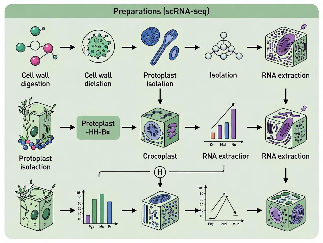
Workflow and Pathway Diagrams
Protoplast to scRNA-seq Workflow
Troubleshooting Low Yield Pathway
The Critical Role of Protoplasts in Accessing Single-Cell Transcriptomes
Single-cell RNA sequencing (scRNA-seq) has revolutionized plant biology by allowing researchers to investigate cellular heterogeneity and gene expression at an unprecedented resolution. Protoplasts, which are plant cells that have had their cell walls enzymatically removed, serve as a primary starting material for these studies. This technical support center addresses the most common challenges and questions researchers face when preparing protoplasts for scRNA-seq, providing troubleshooting guides and detailed protocols to ensure successful experimental outcomes.
Frequently Asked Questions (FAQs)
1. What is the primary advantage of using protoplasts over nuclei for scRNA-seq in plants? Protoplasts provide a more holistic view of the transcriptome because they capture both nuclear and cytoplasmic RNAs. This comprehensive capture allows for a fuller picture of gene expression patterns and regulatory processes within individual cells [4].
2. What is the major drawback of protoplast isolation, and what is a common alternative? A significant drawback is the "transcriptomic shock" or cellular stress induced by the enzymatic digestion process, which can alter gene expression profiles. A common alternative is using isolated nuclei for single-nucleus RNA sequencing (snRNA-seq), which minimizes this stress and can provide better recovery of cell types that are difficult to protoplast [10] [4].
3. Why might my protoplast sample lack certain cell types? Different cell types have varying sensitivities to cell wall-degrading enzymes and possess different wall structures, leading to skewed distributions in the final protoplast suspension. For instance, protoplasting of maize leaves has been known to fail in recovering vascular cells [10].
4. How can I improve the viability of my isolated protoplasts? Viability can be optimized by carefully tuning the enzyme solution. For example, in cabbage, a dramatic decrease in viability (from 97% to 37%) was observed when the concentration of Pectolyase Y-23 was increased from 0.05% to 0.1%. Substituting with 0.1% Macerozyme R-10 resulted in high yields while maintaining viability over 90% [11].
5. Can I use frozen tissue for protoplast isolation? Typically, no. Protoplast isolation generally requires fresh tissue, as the enzymatic digestion process is performed on living cells. In contrast, nuclei can be isolated from frozen or fixed tissue, which is a key advantage of the snRNA-seq approach [10].
Troubleshooting Guides
Problem: Low Protoplast Yield
Potential Causes and Solutions:
- Cause: Suboptimal enzyme concentration or combination.
- Cause: Inadequate digestion time.
- Solution: Optimize the duration of enzymatic digestion. While some tissues like tobacco may digest in 4 hours, others like celery may require 8 hours or more [12].
- Cause: Incorrect osmotic pressure in the enzyme solution.
- Solution: Adjust the concentration of an osmoticum like mannitol. A concentration of 0.6 M was found to be optimal for celery protoplast isolation [12].
Problem: Poor Protoplast Viability
Potential Causes and Solutions:
- Cause: Over-digestion with overly concentrated enzymes, particularly pectinases.
- Solution: Reduce the concentration of pectolytic enzymes. As noted in the cabbage study, high Pectolyase Y-23 concentration (0.1%) severely compromised viability [11].
- Cause: Damage during purification.
- Solution: Use gentle centrifugation speeds. For celery, 200× g was optimal for collecting protoplasts without causing damage [12]. Always use a sucrose or percoll gradient for gentle purification.
- Cause: Oxidative stress after isolation.
- Solution: Include antioxidants in the culture media. Research on cannabis protoplasts has shown that cultured cells exhibit oxidative stress resilience, which is crucial for viability [13].
Problem: Skewed Cell Type Representation in scRNA-seq Data
Potential Causes and Solutions:
- Cause: Inherent bias in the protoplasting efficiency of different cell types.
- Cause: Over-representation of easily digestible cell types like mesophyll.
- Solution: Use mechanical methods to pre-expose harder-to-digest tissues. For example, scraping the bark off poplar stems or using a "tape sandwich" to remove epidermal layers in Arabidopsis leaves can facilitate protoplast release from underlying tissues [10].
Optimized Experimental Protocols
Sample Preparation: Isolation of Leaf Mesophyll Protoplasts
The following table summarizes optimized enzyme solutions for different plant species, demonstrating that protocols must be species-specific.
Table 1: Optimized Enzyme Solutions for Protoplast Isolation from Various Plant Species
| Plant Species | Cellulase Concentration | Pectinase Concentration | Osmoticum (Mannitol) | Digestion Time | Reference |
|---|---|---|---|---|---|
| Cabbage (Brassica oleracea) | 0.5% Cellulase Onozuka RS | 0.1% Macerozyme R-10 | Information Missing | Overnight | [11] |
| Celery (Apium graveolens) | 2.0% Cellulase R-10 | 0.1% Pectolase | 0.6 M | 8 hours | [12] |
| Moss (Physcomitrella patens) | Protocol described | Protocol described | Information Missing | Information Missing | [14] |
| Arabidopsis thaliana | 1.5% Cellulase | 0.4% Pectolase | Information Missing | Information Missing | [12] |
Detailed Protocol for Celery Protoplast Isolation [12]:
- Plant Material: Use leaves from 3-week-old, sterile-grown celery seedlings.
- Tissue Preparation: Slice leaves into very fine strips (0.5–1 mm width) using a sharp blade.
- Enzymatic Digestion: Immerse the leaf strips in the filter-sterilized enzyme solution (2.0% cellulase, 0.1% pectolase, 0.6 M mannitol, pH 5.8).
- Incubation: Incubate in the dark at 25°C with gentle shaking (45 rpm) for 8 hours.
- Purification:
- Filter the resulting protoplast suspension through a 400-mesh sieve to remove undigested debris.
- Centrifuge the filtrate at 200× g to pellet the intact protoplasts.
- Resuspend the pellet in a W5 salt solution (2 mM MES, 154 mM NaCl, 125 mM CaCl₂, 5 mM KCl; pH 5.7).
- Assessment: Count protoplasts using a hemocytometer and assess viability (should be >90%) using FDA staining.
Workflow for scRNA-seq Library Construction
The following diagram illustrates the general workflow for proceeding from plant tissue to scRNA-seq data, highlighting critical steps where troubleshooting is often required.
Method Selection: Protoplasts vs. Nuclei
Choosing between protoplasts and nuclei is a critical first step in experimental design. The following table compares these two primary methods to guide researchers.
Table 2: Comparison of Protoplast-based and Nucleus-based Methods for Plant scRNA-seq
| Feature | Protoplast-based scRNA-seq | Nucleus-based snRNA-seq |
|---|---|---|
| Transcriptome Coverage | Captures nuclear and cytoplasmic RNA (full transcriptome) [4] | Primarily captures nuclear RNA; misses cytoplasmic transcripts [4] |
| Cellular Stress | High ("transcriptomic shock" from 1-2 hour digestion) [10] | Low; minimal perturbation [10] |
| Cell Type Bias | High; some cell types (e.g., vasculature) are difficult to isolate [10] | Lower; better recovery of hard-to-dissociate cell types [4] |
| Sample Flexibility | Requires fresh tissue [10] | Compatible with frozen or fixed tissue [10] [4] |
| Ideal Use Case | Studies requiring full transcriptome data from easily protoplasted tissues (e.g., mesophyll) | Studies of complex tissues, rare cell types, or when sampling requires preservation |
The Scientist's Toolkit: Key Research Reagents
Table 3: Essential Reagents for Protoplast Isolation and scRNA-seq
| Reagent | Function | Example(s) |
|---|---|---|
| Cell Wall-Digesting Enzymes | Break down cellulose (cellulase) and pectin (pectinase) in the plant cell wall. | Cellulase Onozuka RS, Macerozyme R-10, Pectolyase Y-23 [11] [12] |
| Osmoticum | Maintains osmotic pressure to prevent protoplast bursting. | Mannitol (0.4 - 0.7 M) [12] |
| Viability Stain | Assesses the health and integrity of isolated protoplasts. | Fluorescein Diacetate (FDA) [12] |
| Purification Sieve | Removes undigested tissue and debris from the protoplast suspension. | 400-mesh sieve [12] |
| scRNA-seq Library Kit | For barcoding, reverse transcription, and library construction of single-cell transcripts. | 10x Genomics Chromium Single Cell 3' Reagent Kits [14] [4] |
| Protoplast Culture Medium | Supports protoplast regeneration and cell division for downstream applications. | Modified MS or KM media with plant growth regulators [13] |
Successful single-cell transcriptomics in plants hinges on robust protoplast preparation. While challenges such as cellular stress, low yield, and cell type bias persist, they can be overcome with careful optimization of enzymatic cocktails, digestion conditions, and purification methods. By understanding the trade-offs between protoplast and nucleus isolation, and by applying the troubleshooting guidelines presented here, researchers can reliably generate high-quality single-cell suspensions to unlock the cellular heterogeneity of plants.
Frequently Asked Questions (FAQs)
FAQ 1: What are the primary challenges when preparing plant protoplasts for single-cell RNA sequencing (scRNA-seq)?
The three most significant challenges are:
- Cellular Heterogeneity: Tissues consist of many different cell types. A successful protoplast isolation must capture this full diversity without introducing bias towards or against any specific cell type [3] [15].
- Cell Wall Digestion: The plant cell wall is a complex, rigid structure that varies between species, organs, and developmental stages. Incomplete digestion yields low protoplast numbers, while over-digestion can damage cells and induce stress responses, compromising viability and transcriptomic data [16] [1] [17].
- Transcriptional Stress: The protoplast isolation process itself—involving enzymatic digestion, mechanical stress, and changes in osmotic pressure—can trigger rapid and significant changes in gene expression. This makes it difficult to distinguish true biological signals from technical artifacts [3] [1].
FAQ 2: Should I use protoplasts (whole cells) or nuclei for plant scRNA-seq?
The choice depends on your research question and plant material. The table below compares the two approaches.
Table 1: Comparison of Protoplasts vs. Nuclei for scRNA-seq
| Feature | Protoplasts (Whole Cell) | Nuclei (snRNA-seq) |
|---|---|---|
| Transcriptome Coverage | Captures both nuclear and cytoplasmic mRNA, providing a more complete picture of the transcriptome [18]. | Primarily captures nuclear transcripts; may miss some cytoplasmic mRNAs [3] [18]. |
| Applicability | Ideal for tissues with cells that are easily dissociated and have thin walls (e.g., young leaves, roots) [16] [5]. | Essential for tissues that are difficult to dissociate (e.g., woody tissues, fibrous tissues) or when cells are exceptionally large [3] [16] [18]. |
| Stress Response | High risk of inducing transcriptional stress responses during cell wall digestion [16] [1]. | Minimizes stress associated with cell wall digestion, as nuclei can be isolated from frozen tissue, "freezing" the transcriptional state [3] [18]. |
| Spatial Information | Like nuclei, protoplasts lose their native spatial context within the tissue upon isolation [3]. | Loses native spatial context, though spatial transcriptomics techniques can compensate for this [3] [19]. |
FAQ 3: How can I minimize transcriptional stress during protoplast isolation?
Minimizing stress requires a optimized and gentle protocol:
- Optimize Digestion Time: Use the shortest effective digestion time to avoid prolonged stress [20] [5].
- Control Temperature: Perform all steps, especially after digestion, at 4°C to arrest cellular metabolism and reduce stress-related gene expression [18].
- Use Proper Osmotic Stabilizers: Maintain correct osmotic pressure with substances like mannitol or sorbitol to prevent cell lysis or rupture [20] [17].
- Work Quickly: Process samples rapidly from isolation to sequencing or fixation to capture an accurate transcriptional snapshot [18].
FAQ 4: What is a critical step often overlooked in protoplast regeneration for genome editing?
A key step is the inclusion of mycelial extract or other tissue-specific extracts in the regeneration medium. For example, adding Lyophyllum decastes mycelial extract to the Z5 medium significantly increased the regeneration rate to 2.86 [20]. This suggests that species-specific supplements providing essential growth factors can dramatically improve the efficiency of regenerating whole plants from transfected protoplasts.
Troubleshooting Guides
Troubleshooting Low Protoplast Yield and Viability
Table 2: Troubleshooting Protoplast Isolation
| Problem | Potential Cause | Solution |
|---|---|---|
| Low Yield | Inefficient enzyme solution | Systematically optimize enzyme concentrations and combinations (cellulase, macerozyme, pectinase). A two-step digestion process can also improve yield [1]. |
| Suboptimal plant material | Use young, actively growing tissues (e.g., 65-75 hour hydroponically grown cotton roots, 10-day-old fungal mycelia). Older tissues have thicker, more recalcitrant cell walls [20] [5]. | |
| Incomplete tissue digestion | Cut tissue into fine, translucent slices to maximize surface area for enzyme action [5]. | |
| Low Viability (<80%) | Over-digestion | Reduce enzymatic digestion time. Conduct time-lapse experiments to find the optimal window [20] [1]. |
| Osmotic shock | Ensure the osmotic stabilizer (e.g., 0.6M mannitol, MgSO₄) is present in all solutions and is appropriate for your species [20] [5]. | |
| Mechanical damage | Avoid vigorous pipetting or shaking. Use wide-bore pipette tips and round-bottom centrifuge tubes [5] [18]. | |
| High Debris & Clumping | Presence of dead cells and cations | Use calcium- and magnesium-free buffers. Filter the protoplast suspension through a 30-40 μm cell strainer and use density gradient centrifugation to remove debris [19] [18]. |
Addressing Cellular Heterogeneity in scRNA-seq Data
Table 3: Optimizing for Cellular Heterogeneity
| Challenge | Impact on Heterogeneity | Mitigation Strategy |
|---|---|---|
| Size-Based Bias | Droplet-based systems may under-sample large cells [5]. | For large cells, use nuclei (snRNA-seq) or employ a plate-based scRNA-seq platform that is less sensitive to cell size [16] [18]. |
| Rare Cell Types | Rare cell populations may be missed due to insufficient cell numbers [19] [15]. | Sequence a sufficiently high number of cells. For complex tissues, target tens of thousands of cells to ensure rare types are captured [5] [18]. |
| Batch Effects | Technical variation between samples can confound biological differences [19]. | Use combinatorial barcoding to process multiple samples in a single run [15] [18]. Employ batch correction algorithms (e.g., Harmony, Combat) in downstream analysis [19]. |
The Scientist's Toolkit: Essential Reagents and Materials
Table 4: Key Research Reagent Solutions for Protoplast Work
| Reagent / Material | Function | Examples & Notes |
|---|---|---|
| Cellulase R10 | Hydrolyzes cellulose, the primary structural component of the plant cell wall. | A standard enzyme for protoplast isolation; often used at 1.5-2% (w/v) concentration [5] [17]. |
| Macerozyme R10 | A mixture of enzymes that targets pectin in the middle lamella, helping to separate cells. | Typically used at 0.2-0.75% (w/v) [1] [5]. |
| Pectinase | Specifically degrades pectin, crucial for breaking down the cell wall matrix. | Concentration must be optimized; used at 0.5% in some protocols [1]. |
| Osmotic Stabilizer | Prevents protoplast lysis by maintaining osmotic balance. | Mannitol (0.4-0.6 M) is most common. Sorbitol, sucrose, or KCl can also be used [20] [5] [17]. |
| Calcium Chloride (CaCl₂) | Stabilizes the plasma membrane and facilitates protoplast fusion and transfection. | Used in washing solutions (e.g., W5 solution) and to enhance PEG-mediated transfection [5] [17]. |
| Polyethylene Glycol (PEG) | Facilitates the delivery of DNA, RNA, or proteins (like CRISPR RNP) into protoplasts. | PEG 4000 at 40% concentration is standard, but heat shock treatment can further increase efficiency [1] [17]. |
| Miracloth / Cell Strainer | Filters out undigested tissue debris and cell clumps to create a clean protoplast suspension. | Use multiple layers of Miracloth followed by a 30-40 μm nylon mesh strainer [20] [5]. |
Experimental Workflow and Decision Diagrams
Diagram 1: Protoplast Experimental Planning
Diagram 2: Optimal Protoplast Workflow
Within the broader context of troubleshooting plant protoplast preparation for single-cell RNA sequencing (scRNA-seq), a fundamental decision researchers face is the choice of biological starting material. The two primary entities used are protoplasts (whole cells with their walls enzymatically removed) and isolated nuclei (for single-nucleus RNA-seq, or snRNA-seq). This guide details the technical trade-offs between these approaches to help you select the optimal strategy for your experimental goals and sample type, and to troubleshoot common pitfalls associated with plant sample preparation [4] [21].
The following workflow outlines the critical decision points and associated challenges for each path:
Technical Comparison Table
The choice between protoplasts and nuclei involves a direct trade-off between transcriptome completeness and the minimization of technical artifacts. The following table summarizes the core technical differences:
| Feature | Protoplasts | Single Nuclei |
|---|---|---|
| Transcriptome Coverage | Full transcriptome (nuclear + cytoplasmic) [4] | Nuclear transcriptome; loss of cytoplasmic mRNAs [4] |
| Sample Preparation Impact | Enzymatic digestion induces cellular stress & alters gene expression [4] [21] | Minimal perturbation; no cell wall digestion needed [4] [21] |
| Tissue Applicability | Limited by digestibility; recalcitrant tissues (e.g., xylem) are challenging [4] [5] | Broad applicability; suitable for hard-to-digest tissues & frozen samples [4] [21] |
| Cell Size Restrictions | Yes; must be <40-50 µm for droplet-based platforms (e.g., 10x Genomics) [5] | Minimal constraint; nuclei are small and uniform [21] |
| Key Advantage | Captures a more complete picture of cellular gene expression. | Better preserves native transcriptional states; wider tissue applicability. |
| Major Limitation | Stress responses can confound biological interpretations. | Incomplete transcriptome limits analysis of cytoplasmic processes. |
Frequently Asked Questions (FAQs) & Troubleshooting
How do I decide whether to use protoplasts or nuclei for my experiment?
Your choice should be guided by your research question and sample type.
- Choose Protoplasts if: Your biological question involves signaling pathways, metabolic activity, or other processes reliant on cytoplasmic mRNA, and you are working with a tissue that is known to be easily digestible (e.g., young Arabidopsis or cotton roots) [4] [5].
- Choose Nuclei if: You are working with recalcitrant tissues (e.g., woody tissues, xylem), studying nuclear-specific processes like transcription, or are concerned that enzymatic stress will mask your biological signal of interest [4] [21]. Nuclei are also the preferred option when working with archived or frozen samples [4].
I am getting low protoplast yield and viability. How can I improve this?
Low yield and viability are often related to the starting plant material and digestion protocol.
- Optimize Plant Material: The developmental stage is critical. Use youthful and tender tissues. For cotton roots, the optimal window is 65-75 hours after hydroponic culture [5].
- Optimize Enzyme Solution: Fine-tune the concentration of cellulase and macerozyme enzymes. A common starting point is 1.5% Cellulase R10 and 0.75% Macerozyme R10 in an osmoticum like 0.4-0.5 M mannitol [5] [22].
- Control Digestion Conditions: Limit digestion time (typically 3-4 hours) and use gentle shaking (40-50 rpm) to prevent mechanical stress [5].
My protoplasts are clustering during scRNA-seq, suggesting a stress response. How can I mitigate this?
This is a known limitation of protoplasts. To minimize stress:
- Minimize Protocol Duration: Reduce the time from tissue harvesting to protoplast fixation or lysis as much as possible.
- Validate with Controls: Include a "time-zero" control or validate key findings using an independent method, such as spatial transcriptomics or in situ hybridization [21].
- Consider Nuclei: If stress responses remain a persistent problem, switching to a nuclei-based protocol may be necessary to capture a more native transcriptional state [4] [23].
I am not capturing certain cell types in my protoplast/scRNA-seq experiment. What could be wrong?
Some cell types are more resistant to enzymatic digestion or may be physically filtered out.
- Recalcitrant Cell Types: Tissues with thick, lignified secondary cell walls (e.g., xylem vessel elements) are often underrepresented in protoplast preparations [4].
- Size Exclusion: Protoplasts from large cells may be excluded by the mandatory 30-40 µm cell strainer used to prevent microfluidic chip clogging [5].
- Solution: Using nuclei isolation can often recover these missing cell types, as the mechanical homogenization process is less biased by cell wall composition and there is no size-based filtering beyond the initial nuclei purification [4] [21].
The Scientist's Toolkit: Essential Research Reagent Solutions
| Reagent / Material | Function in Protocol | Key Considerations |
|---|---|---|
| Cellulase R10 / Macerozyme R10 | Enzymatic digestion of cellulose and pectin in plant cell walls. | Critical for protoplast yield and viability; concentration and incubation time require optimization for each species and tissue [5]. |
| Mannitol | Osmoticum to maintain osmotic pressure and prevent protoplast bursting. | Standard concentration ranges from 0.4 M to 0.5 M; essential for protoplast integrity [5] [22]. |
| MES Buffer | Maintains stable pH during enzymatic digestion. | Typically used at pH 5.7 to maintain enzyme activity [5]. |
| W5 Solution | Washing and resuspension solution that provides essential ions (Ca²⁺, Na⁺, K⁺). | High Ca²⁺ content helps stabilize protoplast membranes. Note: Ca²⁺/Mg²⁺ must be removed (e.g., by resuspending in mannitol) for some scRNA-seq library preps [5]. |
| Miracloth / Cell Strainers | Filtration to remove undigested tissue debris and cell clumps. | Sequential filtration through 40 µm (or 30 µm) strainers is crucial to obtain a single-cell suspension compatible with microfluidics [5]. |
Optimized Protocols for Protoplast Isolation and scRNA-seq Library Preparation
Protoplast isolation, the process of creating plant cells without cell walls, is a foundational technique for single-cell RNA sequencing (scRNA-seq) in plant biology. This process enables researchers to investigate cellular heterogeneity, gene regulatory networks, and developmental trajectories at an unprecedented resolution. However, the recalcitrance of cell walls, which vary in composition across species, tissues, and developmental stages, presents a significant technical hurdle. This technical support center synthesizes troubleshooting knowledge and optimized protocols from key model species—Arabidopsis thaliana, Brassica, and pea—to help researchers overcome barriers in protoplast preparation for successful scRNA-seq experiments.
Troubleshooting Guides and FAQs
Frequently Asked Questions
Q1: Why is protoplast yield low for my specific plant species? Low yield is often due to suboptimal enzyme combinations or digestion times. The cell wall composition of different species requires tailored enzymatic cocktails. A universal two-step digestion protocol has been successfully applied across diverse angiosperm species. This involves an initial digestion with a pectinase-rich buffer to break down the middle lamella, followed by a secondary digestion focused on cellulase activity to hydrolyze the primary cell wall [1].
Q2: My protoplasts have poor viability. What are the main causes? Poor viability can result from osmotic shock, excessive digestion time, or contamination. Using a pre-treatment buffer with balanced osmotic pressure, vacuum infiltrating for 10 minutes, and strictly controlling digestion time to 3-4 hours can significantly increase viability from approximately 78% to over 90% [1]. Furthermore, filtering protoplasts through 70 µm and 40 µm strainers removes damaging debris [24].
Q3: Does the protoplast isolation process itself alter gene expression? The enzymatic digestion process can induce stress responses. However, studies monitoring key epigenetic regulators found that the protoplasting process did not generate significant transcriptomic fluctuations resulting from epigenetic remodeling, unlike wound responses in intact tissues [1]. Nevertheless, it is recommended to filter out protoplasting-induced transcriptional responses in scRNA-seq data by comparing isolated protoplasts with undigested tissues.
Q4: When should I use nuclei instead of protoplasts for plant scRNA-seq? Single-nucleus RNA sequencing (snRNA-seq) is advantageous when working with tissues that are difficult to digest (e.g., woody species), when protoplasts are too large for microfluidic devices, or when studying processes where spatial information is lost and cannot be recovered. snRNA-seq avoids the stress responses triggered by cell wall digestion and is compatible with frozen or preserved samples [3] [21] [25].
Q5: How can I prevent contamination during the isolation process? Maintaining aseptic technique is critical. Key practices include: using personal protective equipment (PPE) and biosafety cabinets, sanitizing all equipment and gloves with 70% ethanol before use, cleansing the hood after each session, using Plant Preservative Mixture (PPM) in culture media to target contaminants, and minimizing cell exposure to unsterile environments [26].
Troubleshooting Common Problems
Table 1: Common Protoplast Isolation Issues and Solutions
| Problem | Possible Cause | Solution |
|---|---|---|
| Low Yield | Inefficient cell wall digestion | Optimize enzyme ratios (e.g., 1% cellulase, 0.5% pectinase, 0.5% macerozyme for Chirita pumila); implement a two-step digestion protocol [1]. |
| Incomplete tissue digestion | Pre-treat samples with balanced osmotic buffer; cut tissue into small pieces (<1mm) to increase surface area [1] [24]. | |
| Poor Viability | Osmotic shock | Use an appropriate osmoticum (e.g., 0.4-0.5 M mannitol) in all solutions; include MES buffer to maintain stable pH [27] [24]. |
| Enzymatic toxicity | Reduce digestion time; purify protoplasts promptly after digestion by centrifugation and washing [24]. | |
| Cell Clumping | Presence of undigested wall fragments | Filter the protoplast suspension sequentially through 70 µm and 40 µm cell strainers [24]. |
| Excessive debris | Use a wide-bore pipette to handle protoplasts gently and avoid mechanical shear [24]. | |
| Failed Transformation | Low membrane fluidity | Apply a heat-shock treatment (e.g., 45-50°C for 5 min) post-PEG transformation to increase fluidity and DNA uptake [1]. |
| Low-quality protoplasts | Use protoplasts with >80% viability and optimize PEG concentration and exposure time [1]. |
Optimized Experimental Protocols
Arabidopsis thaliana Root Protoplast Isolation for scRNA-seq
This protocol is adapted from a demonstrated method for moss and Arabidopsis root protoplasting, optimized for use with the 10x Genomics Chromium system [24].
Key Reagents:
- Solution A: 0.4 M Mannitol, 20 mM MES (pH 5.7), 20 mM KCl, 10 mM CaCl₂, 0.1% BSA.
- Enzyme Solution: 1.25% Cellulase ("ONOZUKA" R-10), 0.1% Pectolyase in Solution A.
Workflow:
- Plant Growth: Sterilize and sow Arabidopsis seeds densely on nylon-mesh screens placed on solidified MS Growth Media. Grow plates vertically under continuous light at 22°C for 4-5 days until roots are 2-3 cm long.
- Root Harvesting: Decant water from the dish. Use a scalpel to slice off roots and collect them.
- Enzymatic Digestion: Place 4 mL of Enzyme solution in a 35 mm dish with a 70 µm strainer inside. Transfer the harvested roots into the strainer. Digest on a rotating platform at 85 rpm for 45-60 minutes at 25°C, agitating 2-3 times.
- Protoplast Purification: Lift the strainer and collect the liquid. Centrifuge the collected solution at 500g for 10 minutes. Discard the supernatant.
- Filtration and Washing: Gently resuspend the pellet in 500 µL of Solution A (no enzymes). Filter the suspension sequentially through a 70 µm strainer, then two 40 µm strainers.
- Final Concentration: Centrifuge the filtered solution at 200g for 6 minutes. Discard the supernatant and resuspend the protoplast pellet in a small volume (30-50 µL) of Solution A.
- Quality Control: Assess protoplast density, purity, and viability (e.g., using Evans blue staining). Aim for >80% viability and little to no debris. Adjust the concentration to 700-1000 protoplasts/µL for loading on the 10x Genomics Controller.
The entire process from root cutting to loading should not exceed 90 minutes to ensure high-quality transcriptomic data [24].
Universal Two-Step Digestion Protocol for Diverse Species
This protocol, established for Chirita pumila and tested on multiple angiosperm organs, provides a framework that can be adapted for species like Brassica and pea [1].
Key Reagents:
- Pretreatment Buffer: 10 mM MES, 0.47 M Mannitol, 10 mM Calcium (pH 5.8).
- Primary Enzyme Buffer: 1% Cellulase, 0.5% Pectinase, 0.5% Macerozyme in pretreatment buffer.
- Secondary Enzyme Buffer: 1.2% Cellulase, 0.4% Macerozyme.
Workflow:
- Pretreatment: Apply the pretreatment buffer to tissue samples under vacuum infiltration for 10 minutes. This significantly enhances protoplast stability and viability [1].
- Primary Digestion: Incubate pretreated tissues in the Primary Enzyme Buffer for 3-4 hours.
- Secondary Digestion: Replace the solution with the Secondary Enzyme Buffer and incubate for an additional 60-90 minutes. This step effectively dissociates incompletely hydrolyzed tissues and increases yield.
- Protoplast Regeneration: For whole plant regeneration, immobilize protoplasts in a thin alginate layer using a culture density of 1 × 10^6 protoplasts/mL. The entire process from callus formation to de novo root organogenesis can be completed within 15 weeks in optimal conditions for regenerable species like Arabidopsis Wassilewskija (Ws-2) [27].
Figure 1: Universal two-step protoplast isolation workflow.
The Scientist's Toolkit: Research Reagent Solutions
Table 2: Essential Reagents for Protoplast Isolation and Their Functions
| Reagent | Function | Example(s) |
|---|---|---|
| Osmoticum | Maintains osmotic balance to prevent protoplast bursting; provides a stable environment during and after cell wall removal. | Mannitol (0.4-0.5 M), Sucrose (0.6 M) [1] [27] [24]. |
| Cellulase | Degrades cellulose microfibrils, the primary structural component of the plant cell wall. | Cellulase R-10 ("ONOZUKA") [24], Celluclast 1.5 L [27]. |
| Pectinase | Breaks down pectin in the middle lamella, the adhesive between plant cells, enabling tissue dissociation. | Pectolyase [24], Pectinex Ultra SP-L [1]. |
| Macerozyme | A pectinase and hemicellulose-degrading enzyme that macerates plant tissues. | Macerozyme R-10 [1]. |
| Buffer System | Maintains a stable pH throughout the isolation process, which is critical for enzyme activity and cell health. | MES (pH 5.7-5.8) [27] [24]. |
| Calcium Source | Helps maintain membrane integrity and is used for alginate embedding in regeneration protocols. | Calcium Chloride (CaCl₂) [1] [27]. |
| Plant Preservative Mixture (PPM) | A broad-spectrum biocide added to media to prevent microbial contamination (bacteria and fungi) [26]. | PPM [26]. |
Species-Specific Considerations and Protocol Adaptation
Successful protoplast isolation requires careful adaptation of universal principles to the specific biology of the target species.
Figure 2: Protocol adaptation relationships across species.
- Arabidopsis thaliana: As a model organism, it has well-optimized protocols. The ecotype matters; for example, Wassilewskija (Ws-2) has shown higher protoplast regeneration potential than Columbia (Col-0) [27]. Standard root protoplast isolation uses a combination of Cellulase and Pectolyase [24].
- Brassica species: Being closely related to Arabidopsis, protocols are often transferable with minor adjustments. The key is to consider the specific organ (leaf, stem, root) and its developmental stage, as cell wall composition varies accordingly.
- Pea (Pisum sativum): As a lesser-studied species for scRNA-seq, it requires more extensive optimization. Starting with the universal two-step protocol [1] and systematically varying enzyme concentrations and digestion times is recommended. Given its more complex tissue structure, ensuring thorough tissue cutting and vacuum infiltration is crucial for consistent yields.
Mastering species-specific protoplast isolation is a critical step toward democratizing the application of scRNA-seq in plant biology. While foundational protocols from models like Arabidopsis provide an excellent starting point, success with new species hinges on systematic troubleshooting and optimization of key parameters: enzyme cocktails, osmotic stability, and viability maintenance. The growing toolkit, including universal digestion protocols and the alternative of single-nuclei RNA-seq, empowers researchers to tackle an ever-wider array of plant species. These advances will ultimately fuel a deeper understanding of cellular function and regulation across the plant kingdom, with significant implications for crop improvement and fundamental plant science.
In plant single-cell RNA sequencing (scRNA-seq) research, the quality of the starting biological material is paramount. The process begins with the isolation of intact, viable protoplasts, which are plant cells that have had their cell walls removed. The enzymatic cocktail used for cell wall digestion is the most critical factor in this initial step, directly impacting protoplast yield, viability, and the success of downstream scRNA-seq applications. An optimized enzyme solution ensures the efficient release of protoplasts without compromising cellular integrity or introducing stress-induced transcriptional changes that could confound scRNA-seq data interpretation. This guide addresses the common challenges and troubleshooting strategies for optimizing cellulase and macerozyme concentrations—the core components of most protoplast isolation protocols—to generate high-quality protoplasts suitable for sensitive single-cell genomic analyses.
FAQ: Core Concepts for Practitioners
Q1: Why is optimizing cellulase and macerozyme concentration critical for protoplast preparation aimed at scRNA-seq?
The optimization of cellulase and macerozyme is fundamental because these enzymes directly determine the efficiency of cell wall digestion and the physiological state of the resulting protoplasts. For scRNA-seq, the objective is not merely to liberate cells but to do so in a way that preserves their native transcriptional profile. Inadequate digestion, due to low enzyme concentrations, results in low protoplast yield and potential bias towards specific cell types that are more easily released [28]. Conversely, excessive enzyme concentrations or prolonged digestion times can damage the plasma membrane, induce stress responses, and trigger aberrant gene expression, which directly corrupts the transcriptional data obtained from scRNA-seq [17]. Therefore, a balanced optimization is essential to maximize yield and viability while minimizing transcriptional artifacts.
Q2: What are the primary functions of cellulase versus macerozyme in a protoplast isolation cocktail?
The enzymes cellulase and macerozyme have distinct yet complementary roles in breaking down the plant cell wall:
- Cellulase targets cellulose, the primary structural polysaccharide forming the microfibril framework of the cell wall. Its action is crucial for degrading the wall's main load-bearing network [29] [17].
- Macerozyme is a pectinase-rich mixture that primarily degrades pectin, a polysaccharide that acts as the "cement" in the middle lamella, holding adjacent plant cells together [29] [17]. Its action is vital for tissue dissociation and the release of individual cells.
In practice, macerozyme works to separate cells from each other, while cellulase works to remove the remaining wall from each cell. A combination of both is typically required for efficient protoplast isolation [29].
Q3: A standard enzyme cocktail isn't working for my specific plant species. What factors should I consider optimizing?
Plant cell wall composition varies significantly across species, tissues, and growth conditions. If a standard protocol fails, a systematic optimization of the following factors is recommended, as detailed in Table 1:
- Enzyme Concentrations: Titrate the levels of cellulase and macerozyme. Harder tissues often require higher concentrations [28] [17].
- Osmoticum Concentration: The osmotic pressure, maintained by mannitol or sorbitol, is crucial for preventing protoplast lysis or shrinkage [28] [17].
- Enzymatic Digestion Time: The duration of tissue exposure to the enzymes must be balanced to achieve complete digestion without harming the protoplasts [28].
- Tissue Source and Age: Young, actively growing leaves or hypocotyls are generally ideal due to their thinner cell walls [17].
Troubleshooting Guide: Common Issues and Solutions
Problem: Low Protoplast Yield
- Potential Cause 1: Insufficient concentration of cellulase and/or macerozyme.
- Solution: Incrementally increase the enzyme concentrations. For example, in Populus simonii × P. nigra, an optimized system used 2.5% cellulase R-10 and 0.6% macerozyme R-10 [28].
- Potential Cause 2: Inadequate digestion time.
- Potential Cause 3: Suboptimal osmotic pressure.
- Solution: Adjust the concentration of the osmoticum (e.g., mannitol). Test a range from 0.5 M to 0.8 M, as the optimal concentration was found to be 0.8 M for poplar leaves [28].
Problem: Poor Protoplast Viability
- Potential Cause 1: Enzyme concentrations are too high, causing damage to the cell membrane.
- Solution: Reduce the concentration of cellulase and macerozyme and/or shorten the digestion time.
- Potential Cause 2: Osmotic imbalance leads to protoplast bursting or collapse.
- Solution: Verify the osmolarity of all solutions. Ensure the mannitol concentration is correctly prepared and consistent across all steps from digestion to washing and resuspension [17].
- Potential Cause 3: Mechanical stress from rough handling or centrifugation.
Problem: Incomplete Tissue Digestion with Visible Clumps
- Potential Cause 1: Deficiency in pectin-degrading activity.
- Solution: Increase the concentration of macerozyme, which is rich in pectinase. Alternatively, consider supplementing with other pectinase enzymes or pectolyase, as was included at 0.3% in the optimized poplar protocol [28].
- Potential Cause 2: Ineffective enzyme penetration into the tissue.
- Solution: Ensure the plant tissue is finely sliced or chopped to increase the surface area for enzyme action [28].
Optimized Protocol and Data Tables
Detailed Experimental Protocol for Enzyme Optimization
The following procedure, adapted from published studies [28] [29], provides a robust starting point for optimizing protoplast isolation from leaf tissue.
- Plant Material Preparation: Harvest 0.2-0.5 g of young, fully expanded leaves from healthy plants. Using a sharp blade, remove the midrib and slice the leaves into thin strips (0.5-1 mm) to maximize surface area for enzyme contact.
- Enzyme Solution Preparation: Freshly prepare the enzyme solution in a petri dish. A basic, widely applicable formulation includes:
- 1.5% (w/v) Cellulase ONOZUKA R-10
- 0.5% (w/v) Macerozyme R-10
- 0.6 M Mannitol (for osmotic support)
- 10 mM CaCl₂ (for membrane stability)
- 20 mM MES buffer (pH 5.8)
- Filter-sterilize the solution using a 0.45 μm syringe filter.
- Enzymatic Digestion: Transfer the sliced leaf tissue into the enzyme solution, ensuring the tissue is fully immersed. Seal the plate and incubate in the dark with gentle shaking (40-80 rpm) at 25-27°C for 3-6 hours. The optimal time is species-dependent.
- Protoplast Purification:
- After digestion, gently swirl the mixture and filter the suspension through a 40-70 μm nylon mesh into a 50 mL centrifuge tube to remove undigested tissue and debris.
- Centrifuge the filtrate at 100-200 x g for 2-5 minutes to pellet the protoplasts.
- Carefully remove the supernatant and resuspend the pellet in a washing solution (e.g., W5 solution: 125 mM CaCl₂, 5 mM KCl, 154 mM NaCl, 2 mM MES, pH 5.8).
- Repeat the washing step once.
- Yield and Viability Assessment:
- Yield: Count the protoplasts using a hemocytometer under a microscope. Calculate yield as protoplasts per gram of fresh weight tissue (protoplasts/gFW).
- Viability: Mix a small aliquot of protoplasts with an equal volume of 0.4% (w/v) Trypan Blue stain. Viable protoplasts will exclude the dye and remain clear, while dead cells will take up the blue color. Calculate viability as the percentage of unstained protoplasts out of the total counted.
Quantitative Data for Enzyme Optimization
Table 1: Optimized Enzyme Cocktail Formulations from Various Plant Species
| Plant Species | Tissue | Cellulase R-10 (%) | Macerozyme R-10 (%) | Additional Enzymes | Mannitol (M) | Digestion Time (Hours) | Yield (protoplasts/gFW) | Viability (%) |
|---|---|---|---|---|---|---|---|---|
| Populus simonii × P. nigra [28] | Leaf | 2.5 | 0.6 | 0.3% Pectolyase Y-23 | 0.8 | 5 | 2.0 x 10⁷ | >98 |
| Physcomitrium patens [29] | Protonemal tissue | 1.5 | 0.5 | - | 0.5 | 3 | 1.8 x 10⁶* | - |
| Solanum genus (General guidance) [17] | Leaf/Hypocotyl | 1.5 - 2.0 | ~0.5 | (Pectinase often included) | 0.4 - 0.6 | 4 - 6 | Variable | Variable |
Yield for moss is estimated from the protocol description. *Specific concentration for Macerozyme in Solanum not provided; 0.5% is a common standard.
Table 2: Troubleshooting Matrix: Symptoms, Causes, and Adjustments
| Observed Problem | Likely Cause | Recommended Adjustment |
|---|---|---|
| Low yield, intact tissue | Low enzyme activity / concentration | Increase cellulase and macerozyme by 0.5% increments |
| Low yield, tissue macerated | Osmotic imbalance / mechanical stress | Increase mannitol concentration; gentler handling |
| High yield, low viability | Enzyme toxicity / over-digestion | Reduce digestion time or enzyme concentration |
| Cell clumping, few free protoplasts | Insufficient pectin degradation | Increase macerozyme concentration or add pectolyase |
Experimental Workflow and Signaling Pathways
The following diagram illustrates the logical decision-making process for optimizing an enzyme cocktail, from problem identification to solution validation.
The Scientist's Toolkit: Research Reagent Solutions
Table 3: Essential Reagents for Protoplast Isolation and Their Functions
| Reagent | Function / Role in Protoplast Isolation | Example from Literature |
|---|---|---|
| Cellulase ONOZUKA R-10 | Hydrolyzes cellulose, the primary component of the plant cell wall, enabling its breakdown. | Used at 1.5% for moss [29] and 2.5% for poplar [28]. |
| Macerozyme R-10 | Degrades pectins in the middle lamella, dissociating tissues and releasing individual cells. | Used at 0.5% for moss [29] and 0.6% for poplar [28]. |
| Mannitol | Non-penetrating osmoticum; maintains osmotic pressure to prevent protoplast lysis and stabilize them during and after isolation. | Optimal concentration was 0.8 M for poplar [28]; commonly used between 0.5-0.6 M for other species. |
| Calcium Chloride (CaCl₂) | Divalent cations help stabilize the plasma membrane of the newly exposed protoplasts. | Included at 10 mM in the digestion solution for poplar [28] and in wash solutions. |
| MES Buffer | Maintains the optimal acidic pH (typically 5.8) for the activity of the cell wall-degrading enzymes. | Used at 20 mM in the enzyme solution for poplar [28]. |
| Pectolyase Y-23 | A potent pectinase often supplemented to enhance the degradation of tough plant tissues. | Added at 0.3% in the optimized poplar protocol to improve efficiency [28]. |
Step-by-Step Guide to Protoplast Purification and Viability Assessment
This guide provides detailed protocols and troubleshooting advice for the critical steps of protoplast purification and viability assessment. These steps are essential for ensuring the success of downstream applications, particularly single-cell RNA sequencing (scRNA-seq), where the quality and vitality of protoplasts directly impact data quality.
Frequently Asked Questions (FAQs)
Q1: What are the primary methods for purifying protoplasts after enzymatic digestion? The main purification methods are centrifugation-based, each exploiting differences in density:
- Sedimentation (Centrifugal Precipitation): Protoplasts are pelleted at the bottom of a tube. It yields high protoplast numbers but may cause mechanical damage [30].
- Floating Method: Protoplasts float to the surface of a hypertonic sucrose solution during centrifugation, effectively separating them from debris [30].
- Interface Method (Density Gradient Centrifugation): A multi-step gradient of solutions like sucrose or mannitol is used. Viable, intact protoplasts will collect at the interface between solutions of different densities, providing a high-purity fraction [30].
Q2: How can I quickly and accurately assess the viability of my isolated protoplasts? The most common and effective method is fluorescence staining with Fluorescein Diacetate (FDA).
- Principle: Non-fluorescent FDA passively crosses the membrane of living cells. Intracellular esterases hydrolyze FDA into fluorescein, which is fluorescent and accumulates in cells with intact membranes. Dead cells cannot perform this conversion.
- Protocol: Incubate a protoplast sample with FDA (e.g., a final concentration of 0.01-0.1%) for a few minutes. Observe under a fluorescence microscope. Viable cells will show bright green fluorescence, while dead cells will not [1] [31].
- Alternative Probes: Rhodamine 123 can be used to monitor mitochondrial membrane potential and thus cellular activity, providing another indicator of viability [1].
Q3: I am getting a low yield of viable protoplasts. What could be going wrong? Low yield and viability are often linked to the isolation and purification process. Key factors to check are:
- Enzyme Combination and Concentration: The ratio of cellulase, macerozyme, and pectinase is critical and may require optimization for your specific plant species and tissue [1] [30].
- Osmotic Pressure: The osmotic stabilizer concentration must be maintained throughout the entire process to prevent protoplast bursting or shrinkage [1] [17].
- Digestion Time: Over-digestion can damage protoplasts. The optimal duration (often 3-6 hours) must be determined empirically [1] [30].
- Centrifugation Force: Excessive g-force during purification can rupture protoplasts. Use low speeds (e.g., 100 × g for 5-10 minutes) [32].
Q4: My protoplasts appear viable after staining, but they are not dividing in culture. Why? High initial viability does not guarantee regenerative capacity. This issue can stem from:
- Stress from Enzymatic Digestion: The digestion process itself triggers significant transcriptional stress responses that can impair future development [4].
- Epigenetic Changes: Though some studies show key epigenetic regulators remain stable [1], the process may still induce subtle changes affecting cell division.
- Culture Conditions: The osmotic pressure, plant growth regulators, and nutrient composition of the culture media must be meticulously optimized for regeneration, which is often a multi-stage process [32].
Troubleshooting Guide
The following table outlines common problems, their potential causes, and recommended solutions.
| Problem | Potential Cause | Solution |
|---|---|---|
| Low Protoplast Yield | Inefficient cell wall digestion; incorrect enzyme cocktail. | Optimize enzyme types and concentrations (e.g., 1-2% cellulase, 0.5-1% pectinase/macerozyme); use younger, vigorously growing tissues [1] [30] [17]. |
| Low Protoplast Viability | Osmotic shock; over-digestion; mechanical damage. | Maintain consistent osmotic pressure with 0.4-0.6 M mannitol/sorbitol; reduce digestion time; use gentle centrifugation (100-200 × g) [1] [30] [32]. |
| Excessive Cellular Debris | Incomplete filtration; tissue not fully digested. | Filter suspension through a 40-100 μm mesh post-digestion; consider a secondary digestion step for stubborn tissues [1] [30]. |
| Protoplasts Bursting | Hypotonic solution; osmotic imbalance. | Ensure all solutions contain the correct concentration of osmoticum (e.g., mannitol); verify the osmolarity of all buffers [30] [17]. |
| Poor Regeneration | Physiological stress from isolation; suboptimal culture media. | Use a multi-stage media regimen with adjusted plant growth regulators; pre-treat plant material with dark or cold incubation to enhance viability [30] [32]. |
Experimental Protocols
Protocol 1: Purification of Protoplasts via Floating Method
This method is effective for separating viable protoplasts from debris and dead cells.
- Filtered Suspension: After enzymatic digestion, filter the protoplast mixture through a 40-100 μm nylon mesh into a sterile tube to remove undigested tissue clumps [30].
- Centrifuge: Centrifuge the filtrate at a low speed (e.g., 100 × g for 10 minutes) to pellet the protoplasts and debris [32].
- Resuspend: Gently resuspend the pellet in a small volume of W5 solution or a mannitol-based washing solution and place on ice for 30 minutes. During this step, viable protoplasts may rise to the top [32].
- Purify: Carefully collect the band of floated protoplasts from the surface using a pipette.
- Wash: Resuspend the collected protoplasts in a fresh washing solution and centrifuge again at 100 × g for 5 minutes to wash. Repeat if necessary [32].
- Resuspend for Use: Resuspend the final, purified protoplast pellet in an appropriate volume of mannitol-based solution or culture medium for downstream applications.
Protocol 2: Viability Assessment Using Fluorescein Diacetate (FDA)
This is a standard and reliable method for quantifying the percentage of living protoplasts.
- Prepare FDA Stock Solution: Dissolve FDA in acetone to create a stock solution (e.g., 5 mg/mL). This stock can be stored at -20°C.
- Stain Protoplasts: Add the FDA stock solution to a protoplast suspension to achieve a final working concentration of approximately 0.01% (e.g., 2 μL of stock per 1 mL of protoplast suspension). Mix gently and incubate in the dark at room temperature for 5-15 minutes [1] [31].
- Observe Under Microscope: Place a drop of the stained protoplast suspension on a microscope slide.
- Observe using a fluorescence microscope with a blue light excitation filter (typically around 490 nm).
- Viable cells will display bright green fluorescence in their cytoplasm.
- Non-viable cells will remain non-fluorescent.
- Calculate Viability: Count the number of fluorescent (live) and non-fluorescent (dead) cells across several fields of view. Calculate the viability percentage using the formula: Viability (%) = (Number of fluorescent cells / Total number of cells counted) × 100 [1].
Research Reagent Solutions
Essential materials and reagents for protoplast purification and viability assessment are listed below.
| Reagent | Function | Example Usage in Protocol |
|---|---|---|
| Cellulase | Hydrolyzes cellulose, the primary component of the plant cell wall. | Used at 1-2% (w/v) in enzyme solution to break down the cell wall matrix [1] [17]. |
| Macerozyme / Pectinase | Degrades pectin in the middle lamella, separating cells from each other. | Used at 0.5-1% (w/v) in combination with cellulase [1] [32]. |
| Mannitol / Sorbitol | Osmotic stabilizer. Prevents protoplasts from bursting by maintaining osmotic balance. | Used at 0.4-0.6 M in all solutions during isolation, purification, and initial culture [32] [17]. |
| Fluorescein Diacetate (FDA) | Vital fluorescent dye. Converted to fluorescent fluorescein only in living cells. | Used at ~0.01% final concentration for viability staining [1] [31]. |
| Calcium Chloride (CaCl₂) | Stabilizes the plasma membrane and facilitates protoplast fusion. | Commonly included in enzyme and washing solutions (e.g., 1-10 mM) [32] [17]. |
| MES Buffer | Maintains a stable pH during the enzymatic digestion process. | Added to the enzyme solution, typically at 10 mM, pH 5.7 [32]. |
Workflow and Pathway Diagrams
Protoplast Purification and Viability Workflow
This diagram outlines the core workflow from tissue to purified, viable protoplasts, highlighting the three main purification pathways.
Protoplast Viability Assessment Logic
This logic diagram illustrates the decision-making process during Fluorescein Diacetate (FDA) staining, showing how cell physiology determines the staining outcome and final viability count.
Adapting Library Construction Methods (10x Genomics, SMART-seq2) for Plant Protoplasts
Single-cell RNA sequencing (scRNA-seq) represents a transformative technology for investigating cellular heterogeneity, developmental trajectories, and gene regulatory networks in complex biological systems. For plant researchers, adapting established library construction methods like 10x Genomics Chromium and SMART-seq2 to work with protoplasts presents unique technical challenges. This technical support center addresses the specific issues users encounter during plant protoplast preparation and subsequent scRNA-seq library construction, providing targeted troubleshooting guidance framed within the broader context of optimizing plant single-cell research.
Core Concepts and Technical Background
Understanding Your Biological Sample: Protoplasts vs. Nuclei
A fundamental consideration in plant scRNA-seq is whether to work with protoplasts (whole cells without cell walls) or isolated nuclei. Each approach has distinct advantages and limitations that significantly impact experimental outcomes [21].
Protoplast-Based Approaches:
- Provide the complete cellular transcriptome, including both nuclear and cytoplasmic mRNAs [21]
- Can induce artificial stress responses due to the enzymatic digestion required to remove cell walls [21]
- Often suffer from cell-type bias as some plant tissues (especially those with secondary cell walls) are difficult to digest completely [33]
- Compatibility with microfluidic devices can be limited by the large size of some plant protoplasts [21]
Nuclei-Based Approaches (snRNA-seq):
- Bypass challenges associated with cell wall digestion [33]
- Enable work with frozen tissues and difficult-to-dissociate cell types [21]
- Capture only the nuclear transcriptome, potentially missing key biological processes involving cytoplasmic mRNA [21]
- Generally provide fewer unique molecular identifiers (UMIs) and detected genes per biological entity [21]
Table 1: Comparison of Biological Entity Selection for Plant scRNA-seq
| Feature | Protoplasts | Nuclei |
|---|---|---|
| Transcriptome Coverage | Nuclear + cytoplasmic | Primarily nuclear |
| Sample Compatibility | Fresh tissues, limited species | Fresh & frozen tissues, broader species |
| Technical Artifacts | Digestion-induced stress | Potential nuclear RNA leakage |
| Cell Type Bias | High (due to differential digestion) | Lower |
| Protocol Optimization | Species and tissue-specific | More universally applicable |
Technology Selection: 10x Genomics vs. SMART-seq2
The choice between droplet-based (10x Genomics) and full-length (SMART-seq2) scRNA-seq methods involves important trade-offs for plant protoplast research [34].
10x Genomics Chromium (3' end counting):
- Uses microfluidic partitioning to create Gel Beads-in-emulsion (GEMs) containing barcoded oligonucleotides [35]
- Enables high-throughput analysis of thousands of cells simultaneously [36] [35]
- Provides cellular indexing through barcoded beads [35]
- Well-suited for large-scale cell type identification and population studies
SMART-seq2 (Full-length transcript coverage):
- Provides full-length transcript coverage, enabling isoform usage analysis [34]
- Typically processes fewer cells but with greater sequencing depth per cell
- Superior for detecting low-abundance genes and transcript variants [34]
- Requires fluorescence-activated cell sorting (FACS) for cell isolation [34]
Table 2: Technical Comparison of scRNA-seq Methods for Plant Protoplasts
| Parameter | 10x Genomics | SMART-seq2 |
|---|---|---|
| Throughput | High (thousands to millions of cells) [35] | Low to medium (hundreds of cells) |
| Transcript Coverage | 3' end only [35] | Full-length [34] |
| Cell Isolation | Microfluidic partitioning [36] | FACS or manual picking [34] |
| UMI Incorporation | Yes [35] | No [34] |
| Cost per Cell | Lower | Higher |
| Ideal Application | Cell atlas construction, population heterogeneity | Deep transcriptional characterization, isoform analysis |
Frequently Asked Questions (FAQs) and Troubleshooting Guides
Protoplast Preparation and Quality Control
Q1: My protoplast viability is low after isolation. What are the critical factors to improve viability?
Low protoplast viability typically results from issues during cell wall digestion. Key optimization points include:
- Enzyme Composition and Osmolarity: Ensure proper enzyme mixtures (cellulase, pectinase, hemicellulase) and maintain correct osmotic pressure throughout digestion [21]
- Digestion Time: Optimize digestion duration to minimize stress – shorter times may yield healthier protoplasts
- Tissue Age and Type: Younger tissues generally yield more viable protoplasts; adjust protocols for different tissue types [21]
- Temperature: Perform digestion at room temperature or slightly below to reduce metabolic stress
- Handling Technique: Use wide-bore pipettes to minimize shear forces during protoplast manipulation
Q2: I'm observing cell-type bias in my protoplast populations. How can I achieve better representation?
Cell-type bias is common in protoplast studies due to differential sensitivity to digestion [21] [33]. Mitigation strategies include:
- Combined Mechanical and Enzymatic Digestion: Gently disrupt tissues mechanically before enzymatic treatment
- Sequential Digestion: Use shorter digestion cycles with collection of released protoplasts at intervals
- Nuclei Isolation Alternative: Consider switching to nuclei isolation for more uniform cell type representation [33]
- Validation: Use cell-type-specific markers to quantify representation and optimize accordingly
Library Construction and Method Adaptation
Q3: How do I adapt the 10x Genomics Chromium workflow for plant protoplasts, given their larger size and different properties?
Plant protoplasts require specific adaptations for droplet-based systems:
- Protopst Size Filtering: Pre-filter protoplasts to ensure they fall within the compatible size range for microfluidic chips (typically <40μm diameter) [21]
- Loading Concentration Optimization: Due to larger size, you may need to adjust cell loading concentrations; follow manufacturer recommendations but expect to optimize empirically [36]
- Buffer Compatibility: Ensure protoplast resuspension buffers are compatible with the 10x Chemistry – avoid viscous solutions that might clog microfluidic chips
- Pressure Optimization: The 10x Chromium Controller may require pressure adjustments for plant protoplasts [36]
Q4: What are the key considerations when using SMART-seq2 with plant protoplasts?
SMART-seq2 implementation with protoplasts requires attention to:
- Cell Lysis Conditions: Optimize lysis buffer composition to effectively break plant membranes without degrading RNA
- Reverse Transcription Efficiency: Plant protoplasts may contain compounds that inhibit reverse transcription; include appropriate additives
- Amplification Bias: Without UMIs, PCR amplification biases can affect quantification; limit amplification cycles where possible [34]
- Quality Assessment: Use Bioanalyzer or TapeStation to confirm full-length cDNA library quality before sequencing
Technical Challenges and Optimization
Q5: I'm getting high background noise and low gene detection in my plant scRNA-seq data. What could be causing this?
Poor data quality often stems from several potential issues:
- RNA Degradation: Ensure rapid processing after protoplast isolation and use RNase inhibitors
- Excessive Debris: Remove debris through careful filtering (30μm strainers) or gradient centrifugation [37]
- Low Viability: Start with high-viability protoplast preparations (>85% recommended) [37]
- Library Construction Issues: Verify reverse transcription and amplification efficiency through QC steps
- Sequencing Depth: Ensure sufficient sequencing depth – plant genomes may require adjustments to standard recommendations
Q6: How can I handle the high RNA content in chloroplasts and other organelles during plant protoplast scRNA-seq?
Organellar RNA can dominate sequencing libraries in plant protoplast preparations:
- Poly-A Selection: Most scRNA-seq methods use poly-A selection which should exclude non-polyadenylated organellar RNA [37]
- Probe-Based Depletion: Consider using ribosomal RNA depletion kits optimized for plant rRNA
- Bioinformatic Filtering: Remove organellar reads computationally during data processing
- Nuclei Alternative: Isolating nuclei instead of whole protoplasts naturally excludes chloroplast and mitochondrial RNAs [21]
Experimental Workflows and Protocol Guidance
Decision Framework for Plant Protoplast scRNA-seq
The following workflow diagram outlines the key decision points when designing a plant protoplast scRNA-seq experiment:
Comprehensive Protocol: 10x Genomics Chromium Adaptation for Plant Protoplasts
Sample Preparation through Library Construction
Protoplast Isolation (Day 1, 3-4 hours)
- Harvest plant tissue and quickly chop into fine pieces in enzyme solution
- Digest with optimized enzyme mixture (concentration and time vary by species/tissue)
- Filter through 30-40μm mesh to remove undigested debris [37]
- Purify protoplasts by centrifugation through sucrose or Percoll gradient
- Resuspend in appropriate buffer and count using hemocytometer
- Assess viability (>85% recommended) using fluorescent dyes [37]
Quality Control Checkpoints
- Microscopy: Examine protoplast morphology – avoid granular or burst protoplasts
- Concentration: Adjust to 700-1,200 cells/μL targeting 10,000 cells per channel (adjust for recovery expectations) [36]
- Viability Re-check: Confirm viability immediately before loading onto Chromium chip
10x Genomics Library Preparation (Day 1, ~1 hour hands-on)
- Use Chromium Single Cell 3' Reagent Kits following manufacturer protocols with modifications:
- Chip Loading: Load protoplast suspension carefully, accounting for larger size
- Partitioning: Run on Chromium Controller or Chromium X Series [36] [35]
- GEM Generation: Monitor GEM formation – plant protoplasts may require pressure adjustments
- Reverse Transcription: Proceed immediately after partitioning [35]
Post-RT Processing and Library Construction (Day 2, 4-5 hours)
- Break emulsions and purify cDNA
- Amplify cDNA with appropriate cycle number (potential optimization point)
- Fragment and size select cDNA before library construction
- Incorporate sample indices and perform final library QC
Sequencing and Data Analysis
- Sequence on Illumina platform with recommended read lengths (28bp Read1, 91bp Read2, 10bp I7 Index)
- Process data through Cell Ranger pipeline with custom reference if needed
- Visualize and explore data with Loupe Browser [35]
The Scientist's Toolkit: Essential Research Reagents and Materials
Table 3: Key Research Reagent Solutions for Plant Protoplast scRNA-seq
| Reagent/Category | Specific Examples | Function/Application | Technical Notes |
|---|---|---|---|
| Cell Wall Digestion Enzymes | Cellulase, Pectinase, Macerozyme | Protoplast isolation from plant tissues | Concentration must be optimized for each tissue type [21] |
| Osmotic Stabilizers | Mannitol, Sorbitol, KCl | Maintain protoplast integrity during isolation | Concentration typically 0.4-0.6M depending on species |
| Viability Stains | Fluorescein diacetate (FDA), Propidium iodide | Assess protoplast viability and membrane integrity | >85% viability recommended for scRNA-seq [37] |
| RNase Inhibitors | Protector RNase Inhibitor, SUPERase-In | Prevent RNA degradation during processing | Critical during protoplast isolation and lysis |
| 10x Genomics Kits | Single Cell 3' Reagent Kits | Library construction for 3' end counting | Compatible with fresh protoplasts; may require optimization [36] |
| SMART-seq2 Reagents | Template Switching Oligo, SMARTER Oligos | Full-length cDNA synthesis and amplification | Enables isoform-level analysis [34] |
| mRNA Capture Beads | Oligo(dT) magnetic beads | mRNA purification for SMART-seq2 | Alternative to poly-A selection in droplets |
| Microfluidic Chips | 10x Genomics Single Cell Chips | Partitioning cells for barcoding | Single-use only to avoid cross-contamination [37] |
| Nuclei Isolation Kits | GEXSCOPE Nuclei Isolation Kit | Nuclear transcriptome preparation | Alternative to protoplasts [37] |
Advanced Troubleshooting: Addressing Complex Challenges
Multi-Omic Integration and Specialized Applications
Integrating snRNA-seq with ATAC-seq: For studies requiring chromatin accessibility data alongside transcriptomics, nuclei isolation enables multi-omic approaches. Isolated nuclei can be split for simultaneous snRNA-seq and snATAC-seq, providing complementary regulatory information [21].
Working with Challenging Tissues: For lignified tissues, glandular structures, or specific cell types resistant to protoplast isolation, the nuclei isolation approach provides a valuable alternative [33]. The Populus protocol demonstrates successful snRNA-seq from shoot apices and stems with varying lignification levels [33].
Cross-Platform Validation: When adopting new methods or troubleshooting existing protocols, consider running a small pilot study comparing protoplast and nuclei approaches from the same tissue source. This validation can help identify method-specific biases and confirm biological findings across technical platforms.
Successfully adapting library construction methods for plant protoplasts requires careful consideration of both biological and technical factors. The protoplast versus nuclei decision represents the most fundamental choice, with implications throughout the experimental workflow. As plant single-cell technologies continue to evolve, methodologies are likely to improve with enhanced protoplast isolation techniques, more sensitive library preparation methods, and integrated multi-omic approaches. By systematically addressing the troubleshooting points outlined in this guide and applying the appropriate experimental frameworks, researchers can overcome the unique challenges of plant protoplast scRNA-seq and unlock deeper insights into plant biology at cellular resolution.
Solving Common Protoplast Preparation Problems for High-Quality scRNA-seq Data
Preventing RNA Degradation During Protoplast Isolation
In single-cell RNA sequencing (scRNA-seq) research, the integrity of RNA is paramount for capturing an accurate snapshot of cellular gene expression. The very process of protoplast isolation—which requires enzymatic digestion of the rigid plant cell wall—poses a significant risk of RNA degradation. This guide provides targeted troubleshooting strategies to help researchers maintain RNA integrity, ensuring that subsequent transcriptomic analyses reflect the true biological state of the cell.
FAQ: Core Challenges in Protoplast RNA Integrity
Q1: Why is protoplast isolation particularly risky for RNA quality? The isolation process subjects plant cells to multiple stressors. Enzymatic digestion buffers can induce global transcriptional changes related to stress responses [1] [38]. Furthermore, the physical breakdown of the cell wall and subsequent handling can activate endogenous RNases. Unlike bulk RNA-seq, where minor degradation might be averaged out, scRNA-seq is exceptionally sensitive to RNA quality drops in individual cells, which can obscure rare cell types and skew developmental trajectories [21] [4].
Q2: What are the critical control points during protoplast isolation? The key control points are: Sample Pretreatment (to minimize cellular stress), Enzymatic Digestion (optimizing conditions to reduce transcriptional artifacts), and Post-Isolation Handling (preventing introduced RNase contamination and further stress) [1] [5]. Maintaining a cold environment and using RNase-free reagents from this point onward is non-negotiable.
Q3: How can I quickly assess the success of my RNA preservation? Beyond standard bioanalyzer measurements, the viability of the protoplasts themselves is a strong proxy. Protoplast viability exceeding 80-90%, as measured by fluorescein diacetate (FDA) or similar staining, is a strong indicator of healthy cells with a high likelihood of intact RNA, and is a common requirement for platforms like the 10x Genomics Chromium system [1] [5].
Troubleshooting Guide: Common Problems and Solutions
Table 1: Troubleshooting RNA Degradation During Protoplast Isolation
| Problem | Potential Cause | Recommended Solution |
|---|---|---|
| Low RNA yield & degradation | Endogenous RNase activation during tissue disruption | Homogenize samples in short bursts (30-45s) with 30s rest to avoid heat buildup; add beta-mercaptoethanol (2-ME) to lysis buffer to inactivate RNases [39] |
| Low RNA yield & degradation | Improper sample storage or handling | Flash-freeze tissue in liquid nitrogen immediately after collection; store at -80°C; keep protoplasts on ice during all post-isolation steps [39] |
| Low RNA yield & degradation | Over-digestion with cell wall enzymes | Systematically optimize enzyme concentrations and incubation time; use a two-step digestion to increase yield without excessive stress [1] [38] |
| DNA contamination | Genomic DNA not removed from sample | Perform an on-column or in-tube DNase treatment during the RNA extraction process [39] |
| Clogged columns during RNA extraction | Incomplete tissue disruption or too much starting material | Increase homogenization time; centrifuge after digestion to pellet debris; transfer only supernatant to column; reduce amount of starting material [39] |
| Stressed cell transcriptome | Osmotic imbalance or chemical stress from enzymes | Use a pre-plasmolysis step with a balanced osmoticum (e.g., mannitol); vacuum infiltrate pretreatment buffer [1] [5] |
Optimized Experimental Protocol for scRNA-seq
The following protocol, adapted from established methods in Chirita pumila and cotton, is designed to maximize protoplast yield and viability while minimizing RNA degradation [1] [5].
Sample Preparation and Pretreatment
- Plant Material: Use young, tender tissues. For roots, 65-75 hours of hydroponic growth after germination is optimal in cotton [5].
- Plasmolysis: Slice tissue into fine, 0.5-1 mm strips and immerse in a plasmolysis solution (e.g., CPW solution with 0.4-0.5 M mannitol) for 30 minutes. This initial osmotic balance helps precondition the cells [1].
- Vacuum Infiltration: Submerge the sliced tissue in an appropriate enzyme solution and apply a gentle vacuum for ~10 minutes. This significantly increases protoplast stability and viability [1] [38].
Enzymatic Digestion
- Enzyme Solution: A common effective mixture includes Cellulase R-10 (1.0-1.5%), Macerozyme R-10 (0.4-0.75%), and Pectinase (0.5%) in an osmoticum (e.g., 0.4-0.5 M mannitol) buffered with MES (pH 5.7) [1] [5].
- Tip: Pre-warm the enzyme solution to 55°C for 10 minutes, then cool to room temperature before filtering and adding CaCl₂ (to 10 mM) and BSA (0.1%) for stability [5].
- Digestion: Incubate in the dark with gentle shaking (40-50 rpm) for 3-5 hours. A two-step digestion protocol (primary digestion followed by a secondary digestion with a modified enzyme mix) can further boost yield from recalcitrant tissues [1].
- Stop Digestion: Add an equal volume of cold W5 solution (154 mM NaCl, 125 mM CaCl₂, 5 mM KCl, 2 mM MES pH 5.7) to halt the enzymatic reaction [5].
Protoplast Purification and RNA Extraction
- Filtration and Washing: Filter the protoplast suspension through 30-40 μm nylon mesh to remove undigested debris. Wash the protoplasts by centrifuging at 100 g for 5-10 minutes in a swinging-bucket rotor and resuspending in cold W5 solution [5].
- Viability Check: Assess viability using FDA staining. Target viability should be >90% for high-quality scRNA-seq [1].
- RNA Extraction: While specific lysis methods depend on the extraction kit, universal best practices include:
- Working quickly on ice.
- Using guaranteed RNase-free tubes and tips.
- Adding β-mercaptoethanol to lysis buffers if not already present.
- Optional: Including a DNase I digestion step to remove genomic DNA contamination [39].
Diagram: Protoplast Isolation Workflow for High-Quality RNA. This workflow highlights the critical steps where RNA is most vulnerable and outlines key protective actions.
The Scientist's Toolkit: Essential Reagents
Table 2: Key Reagents for RNA-Integrity-Focused Protoplast Isolation
| Reagent/Category | Example | Function & Importance |
|---|---|---|
| Osmoticum | Mannitol (0.4-0.6 M) | Maintains osmotic balance to prevent protoplast bursting, a primary stressor. |
| Cell Wall Digestion Enzymes | Cellulase R-10, Macerozyme R-10, Pectinase | Hydrolyzes cellulose, hemicellulose, and pectin. Concentration and ratio must be optimized per species/tissue [1] [40]. |
| RNase Inhibitors | β-Mercaptoethanol, RNase Erase | Critical for inactivating RNases released during tissue disruption. β-ME is added to lysis buffers, while surfaces are treated with specialized solutions [39]. |
| Wash & Resuspension Buffers | W5 Solution, Mg²⁺-free MMG | Used to stop digestion and wash protoplasts. Ca²⁺ helps maintain membrane integrity. For scRNA-seq, mannitol is often preferred over buffers with Ca²⁺/Mg²⁺ that can interfere with reverse transcription [5]. |
| Viability Stain | Fluorescein Diacetate (FDA) | A rapid, reliable method to assess protoplast health and, by proxy, the likelihood of intact RNA before proceeding to costly scRNA-seq [1]. |
Preventing RNA degradation during protoplast isolation is not a single step but an integrated practice spanning experimental design, careful execution, and rigorous quality control. By understanding the stressors introduced at each stage—from the initial slice of the tissue to the final protoplast suspension—researchers can systematically implement the strategies outlined here. Success hinges on optimizing the digestion to minimize cellular stress, relentlessly inhibiting RNases, and verifying protoplast viability. Mastering this foundation is essential for generating robust, high-resolution single-cell transcriptomic data that can power discoveries in plant development and stress biology.
Within the framework of troubleshooting plant protoplast preparation for single-cell RNA sequencing (scRNA-seq), the optimization of culture conditions is paramount. The preparation of viable, high-quality protoplasts is a critical first step, and this process is profoundly influenced by the hormonal environment of the source tissue and the isolation protocol. This guide addresses the pivotal role of plant growth regulators, specifically auxins and cytokinins, in establishing a cellular context conducive to successful protoplast isolation and subsequent scRNA-seq analysis. It provides a targeted FAQ and troubleshooting resource for researchers navigating the technical challenges in this specialized field.
Frequently Asked Questions (FAQs)
1. Why are auxin and cytokinin levels relevant to protoplast isolation for scRNA-seq? Auxin and cytokinin are central regulators of cell division, proliferation, and cellular plasticity [41] [42]. Their balance determines the developmental state and metabolic activity of plant cells. For protoplast isolation, tissue with high meristematic activity (often promoted by a specific auxin:cytokinin ratio) typically contains cells with less rigid walls and higher division potential, which can lead to more efficient protoplast release and higher subsequent viability [42] [43]. Furthermore, these hormones regulate the expression of key genes involved in cell wall remodeling and stress responses, which directly impacts the success of enzymatic digestion and protoplast health [44].
2. How can protoplast isolation itself affect the hormonal transcriptome? Protoplast isolation is a stressful process that involves enzymatic digestion of the cell wall and can significantly alter gene expression. Bulk RNA-seq studies in cotton roots have shown that the isolation procedure can change the expression profile of hundreds of genes, including many involved in plant hormone signal transduction pathways such as auxin and ABA [43]. This means the very act of protoplast preparation can induce a stress response that masks the native transcriptional state of the cell. Therefore, optimizing isolation to minimize this disruption is crucial for obtaining biologically relevant scRNA-seq data.
3. What is a key limitation of scRNA-seq that relates to hormone signaling? A major limitation of standard scRNA-seq protocols is the loss of spatial information. During tissue dissociation into protoplasts, the original location of each cell is lost [45]. Since hormone signaling often operates through precise local gradients and cell-to-cell communication—for instance, auxin maxima and minima that pattern organs—this loss of context can make it difficult to interpret the role of hormone-related genes identified in the scRNA-seq data [41] [45]. Integrating scRNA-seq with spatial transcriptomics techniques is a promising strategy to overcome this limitation.
Troubleshooting Guide: Protoplast Preparation and Hormonal Context
The following table outlines common problems, their potential causes related to growth regulators and culture conditions, and recommended solutions.
| Problem | Possible Hormonal/Condition-Related Cause | Proposed Solution |
|---|---|---|
| Low protoplast viability | Tissue source is senescing or has low cellular activity; incorrect hormone pre-treatment. | Use young, meristematic tissues (e.g., root tips, shoot apex). Pre-culture donor plants or explants on medium with balanced auxin/cytokinin to boost cell activity [42] [43]. |
| Poor protoplast yield | Cell walls are too rigid or lignified, often due to the tissue's developmental state. | Optimize the hormonal pre-conditioning of source plants to maintain cells in a more juvenile state. Systematically optimize enzyme concentration and digestion time [43]. |
| High levels of stress gene expression | The isolation procedure is overly harsh, inducing a strong wounding and stress response. | Shorten the enzymatic digestion time. Incorporate antioxidants into the enzyme and washing solutions. Validate protocol by comparing transcriptomes before/after dissociation [43]. |
| Failure to regenerate cell wall or divide post-isolation | The protoplasts lack the hormonal signals or cellular plasticity to re-enter the growth cycle. | Culture isolated protoplasts in a medium containing a balanced ratio of auxin and cytokinin, which is critical for initiating cell division and de novo organogenesis [42]. |
| Inconsistent results between batches | Uncontrolled variation in the physiological state of the source plant material. | Standardize the growth conditions, age, and harvesting time of the plant material. Pre-condition plants under identical environmental regimes before protoplast isolation. |
Key Experimental Protocols and Data
Optimizing Protoplast Isolation from Root Tips
A study on cotton (Gossypium arboreum) provides a quantitative framework for optimizing protoplast isolation, a methodology that can be adapted for other species. Key parameters were systematically tested to achieve high yield and viability suitable for scRNA-seq [43].
Detailed Methodology:
- Plant Material: 5-day-old root tips were identified as the optimal tissue, yielding protoplasts with over 85% viability [43].
- Vacuum Infiltration: A pressure of 0.05 MPa for 1 hour was optimal for facilitating the infiltration of the enzymolysis solution without damaging the cells [43].
- Enzymatic Digestion: A 6-hour digestion period yielded the highest number of protoplasts (2.00 × 10^6 protoplasts per gram fresh weight) while maintaining sufficient viability [43].
Summary of Optimization Data:
| Parameter Tested | Optimal Condition | Resulting Outcome |
|---|---|---|
| Tissue Age | 5-day-old root tips | Highest yield and >85% viability |
| Vacuum Treatment | 1 hour at 0.05 MPa | Best balance of yield and cell integrity |
| Enzymatic Digestion Time | 6 hours | Peak yield of 2.00 × 10^6 protoplasts/g FW |
The Molecular Interplay of Auxin and Cytokinin in Cell Reprogramming
Understanding the molecular pathways governed by auxin and cytokinin is essential for troubleshooting, as their signaling directly impacts the success of protocols involving protoplasts and regeneration.
- Auxin Signaling: Auxin promotes dedifferentiation and callus formation. It acts through the TIR1/AFB receptors, leading to the degradation of Aux/IAA repressors and the activation of Auxin Response Factors (ARFs). These ARFs in turn regulate genes involved in cellular reprogramming, such as LEC1 and LEC2 [42].
- Cytokinin Signaling: Cytokinin counteracts auxin and promotes cell division. Its signaling is mediated by the Arabidopsis Response Regulator (ARR) family. A key interaction is the cytokinin-mediated promotion of WUSCHEL (WUS), a gene critical for maintaining stem cell identity in the shoot apical meristem [42].
- The Regulatory Network: The balance between these two hormones dictates developmental outcomes. High auxin-to-cytokinin ratios favor root formation, while low ratios promote shoot initiation. This balance is crucial for re-establishing growth from isolated protoplasts [42].
Signaling Pathways and Experimental Workflows
Hormonal Regulation of Cell Reprogramming
This diagram illustrates the core signaling pathways of auxin and cytokinin and their convergence on key genes that control cell fate during de novo organogenesis, a process relevant to protoplast regeneration.
Protoplast Isolation for scRNA-seq Workflow
This flowchart outlines the key steps in a standard protoplast isolation workflow for scRNA-seq, highlighting critical decision and optimization points that impact the quality of the final data.
The Scientist's Toolkit: Research Reagent Solutions
The following table details essential materials and their functions in protoplast-based research for scRNA-seq.
| Research Reagent | Function in Experiment | Specific Example / Note |
|---|---|---|
| Cell Wall Digesting Enzymes | Degrades cellulose and pectin to release protoplasts from tissue. | Macerozyme and Cellulase; concentration and combination must be optimized for each species and tissue type [43]. |
| Fluorescence-Activated Cell Sorter (FACS) | Isolates and purifies specific cell types or nuclei from a heterogeneous protoplast/nuclei suspension. | Can be used to select for viable cells or to reduce background in snRNA-seq [46] [4]. |
| 10x Genomics Chromium Kit | A widely used, high-throughput platform for constructing barcoded scRNA-seq libraries from thousands of single cells. | Commonly applied in plant research using both protoplast and nuclei suspensions [41] [4]. |
| Plant Growth Regulators (Auxins/Cytokinins) | Pre-condition the physiological state of source tissue to improve protoplast yield and viability. | Auxins (e.g., IAA, NAA) and Cytokinins (e.g., BAP) are used in pre-culture media [42]. |
| Triphenyltetrazolium Chloride (TTC) or FDA | Stains metabolically active cells to assess protoplast viability before proceeding to sequencing. | A crucial QC step; aim for >85% viability for optimal results [43]. |
| Polyethylene Glycol (PEG) | Used to induce protoplast fusion for creating novel somatic hybrids or for transfection. | Fusion can disrupt cellular physiology and should be accounted for in experimental design [44]. |
Plant single-cell RNA sequencing (scRNA-seq) represents a transformative approach for investigating cellular heterogeneity, developmental trajectories, and stress responses at unprecedented resolution. However, the path to high-quality data is often obstructed by technical challenges, particularly when working with difficult-to-digest tissues such as lignified cells and root samples. The inherent structural complexity of plant cell walls, varying degrees of lignification across cell types, and the delicate nature of root tissues necessitate optimized and tailored approaches for successful protoplast isolation. This technical support center addresses these specific challenges through targeted troubleshooting guides and frequently asked questions, providing researchers with practical methodologies to overcome common obstacles in protoplast preparation for scRNA-seq.
FAQs and Troubleshooting Guides
FAQ 1: What are the primary challenges when preparing protoplasts from lignified root tissues, and how can they be overcome?
Answer: Lignified tissues present three primary challenges: robust cell walls resistant to enzymatic digestion, potential loss of rare cell types during aggressive processing, and altered gene expression due to protoplasting stress.
Challenge 1: Resilient Cell Walls. Lignin and other complex polymers in secondary cell walls create a physical barrier that is difficult for enzymes to penetrate. Standard cellulase treatments are often insufficient.
- Solution: Implement a tailored enzymatic cocktail. Research on soil-grown roots suggests using combinations of cellulase, pectolyase, and other targeted enzymes. For instance, a demonstrated protocol for Arabidopsis roots uses 1.25% cellulase ("ONOZUKA" R-10) combined with 0.1% pectolyase to effectively break down root tissue [24]. For more fibrous tissues, collagenase (Type I or II) or hyaluronidase can be added to digest specific extracellular matrix components [47].
Challenge 2: Cellular Heterogeneity and Rare Cells. Harsh digestion conditions can preferentially damage or destroy certain delicate cell types, skewing the resulting transcriptome data.
- Solution: Consider single-nucleus RNA sequencing (snRNA-seq) as an alternative. This approach is often a safer compromise for challenging tissues, as isolating nuclei is quicker and performed at colder temperatures, minimizing stress responses and preserving rare cells that might be lost during tissue digestion [3] [47]. This is particularly relevant for root hair cells, which are easily lost during protoplasting of soil-grown roots [48].
Challenge 3: Protoplasting-Induced Stress. The enzymatic digestion and mechanical disruption process itself can trigger rapid gene expression changes, confounding biological interpretations.
- Solution: Minimize processing time and work at cold temperatures. It is recommended to keep the time between root cutting and loading protoplasts onto the sequencer to under 90 minutes [24]. Lower temperatures slow down RNA degradation and stress responses. Furthermore, researchers can identify and bioinformatically exclude genes induced by the protoplasting process from downstream analyses to ensure robustness [48].
FAQ 2: How does growth medium (e.g., soil vs. gel) impact root transcriptomes and protoplasting efficiency?
Answer: The growth medium fundamentally influences root biology and, consequently, the experimental workflow and outcomes. Soil-grown roots exhibit significant transcriptional and morphological differences compared to gel-grown roots, which directly impacts protoplasting.
Transcriptional Differences: Single-cell transcriptomics has revealed that soil-grown roots undergo major expression changes, particularly in outer root cell types (epidermis, exodermis, sclerenchyma, and cortex). These changes are related to nutrient homeostasis, cell wall integrity, and defence responses when compared to homogeneous gel conditions [48]. This means the baseline gene expression profile of your cells is medium-dependent.
Cell Wall Composition: The adaptation to a heterogeneous soil environment likely involves remodeling of cell wall architecture, which can alter the resistance of different cell types to enzymatic digestion. This necessitates potential optimization of digestion protocols for soil-derived samples.
Physical Considerations: Soil-grown roots may have more debris, associated microorganisms, and physical damage that can complicate protoplast isolation and viability. Additional washing steps and careful filtering are essential. Multiple filtering steps through 70 µm and 40 µm strainers are critical to remove debris before loading cells onto a droplet-based system [24].
FAQ 3: What steps can be taken to maximize cell viability and yield during isolation?
Answer: High viability and yield are critical for capturing the full spectrum of cellular diversity. Key considerations include:
Optimized Dissociation Method: Use a balanced combination of mechanical and enzymatic dissociation. Gentle mechanical homogenization (e.g., using a gentleMACS Dissociator or gentle pipette trituration) can be combined with a tailored enzymatic cocktail to improve efficiency without excessive damage [47].
Temperature Control: Perform digestion steps at lower temperatures where possible (e.g., 25°C as in the Arabidopsis protocol [24]) to slow metabolic processes that lead to RNA degradation and cell death, even if enzymatic activity is slower than at 37°C.
Viability Assessment: Always assess viability and cell integrity before proceeding to sequencing. Using dyes like trypan blue or more accurate fluorescent dyes like propidium iodide (PI) allows for a precise viability check under a microscope [47] [24]. A viability of >80% is recommended [24].
The table below summarizes common problems and their solutions for protoplasting difficult plant tissues.
Table 1: Troubleshooting Guide for Protoplast Isolation from Difficult Tissues
| Problem | Potential Cause | Solution |
|---|---|---|
| Low cell yield | Incomplete tissue digestion; over-aggressive filtering | Optimize enzyme concentration and incubation time; use wide-bore pipettes and sequential filtering [24] |
| Low cell viability | Over-digestion; harsh mechanical disruption; prolonged processing | Shorten enzymatic incubation; use gentler mechanical methods; minimize time from harvest to fixation [47] [24] |
| High debris in suspension | Inadequate filtration; tissue damage during collection | Use multiple filtration steps (e.g., 70µm followed by 40µm) [24] |
| Clogging of microfluidic channels | Cell aggregates; large cells; debris | Ensure complete dissociation; filter aggressively; for large cells (>30µm), consider snRNA-seq or combinatorial barcoding [47] |
| Loss of specific cell types | Differential sensitivity to digestion | Validate with marker genes; consider snRNA-seq to preserve rare types [48] [47] |
Experimental Protocols
Detailed Methodology: Protoplast Preparation from Arabidopsis Roots
This protocol is adapted from a demonstrated method for preparing root protoplasts for 10x Genomics scRNA-seq [24].
1. Materials and Reagents
- MS Growth Media: 0.3% Gelrite, 0.2g/L MES, 1% sucrose, 4.33g/L MS salts, pH to 5.7-5.8 with KOH.
- Solution A: 0.4M Mannitol, 20mM MES (pH 5.7), 20mM KCl, 10mM CaCl2, 0.1% BSA.
- Enzyme Solution: To 5ml of Solution A, add 1.25% Cellulase (“ONOZUKA” R-10) and 0.1% Pectolyase.
2. Step-by-Step Procedure
- Plant Growth: Sterilize Arabidopsis seeds and sow densely on nylon-mesh screens placed on MS Growth Media in square Petri dishes. Orient plates vertically and grow under continuous light at 22°C for 4-5 days until roots are 2-3 cm long.
- Tissue Harvest: Open the dish and decant water. Use a scalpel to slice off roots and scrape them into a 70 µm strainer sitting in a 35 mm Petri dish containing 4 ml of Enzyme Solution.
- Tissue Digestion: Place the dish on a rotating platform (85 rpm) for 45-60 minutes at 25°C. Agitate the roots gently with forceps 2-3 times during digestion.
- Protoplast Collection: Lift the strainer out of the solution. Use a wide-bore transfer pipette to collect the liquid from the dish and outside of the strainer. Transfer to a 15 ml tube.
- Centrifugation: Centrifuge at 500g for 10 minutes at 22°C. Carefully discard the supernatant.
- Resuspension and Filtration: Gently resuspend the pellet in 500 µl of Solution A (no enzymes). Filter the suspension sequentially through a 70 µm strainer, then two 40 µm strainers, collecting the liquid from the outside of each strainer.
- Final Purification: Centrifuge the filtered solution at 200g for 6 minutes. Discard the supernatant and gently resuspend the purified protoplasts in 30-50 µl of Solution A.
- Quality Control: Examine protoplasts under a microscope to assess density and viability (e.g., with Evans blue stain). Adjust the concentration to 700-1,000 protoplasts/µl with Solution A for loading onto the 10x Genomics Controller.
The following diagram illustrates the key steps and decision points in this protocol:
The Scientist's Toolkit: Research Reagent Solutions
The table below lists essential reagents and their functions for successful protoplast isolation from difficult plant tissues.
Table 2: Essential Reagents for Plant Protoplast Isolation for scRNA-seq
| Reagent | Function | Example / Note |
|---|---|---|
| Mannitol | Osmoticum to maintain protoplast stability and prevent bursting. | Used at 0.4M in Solution A [24]. |
| Cellulase (e.g., "ONOZUKA" R-10) | Breaks down cellulose microfibrils in the primary cell wall. | A core component of most digestion cocktails [24]. |
| Pectolyase | Degrades pectin, a component of the middle lamella that holds cells together. | Critical for tissue dissociation; used at lower concentrations (0.1%) [24]. |
| MES Buffer | Maintains a stable pH during the digestion process. | pH is typically adjusted to 5.7-5.8 [24]. |
| BSA (Bovine Serum Albumin) | Acts as a protein stabilizer and can reduce adhesion and aggregation of protoplasts. | Included in Solution A at 0.1% [24]. |
| Calcium Chloride (CaCl2) | Helps maintain membrane integrity and stability of the isolated protoplasts. | Used at 10mM in Solution A [24]. |
| Collagenase | Digests collagen-like proteins in the extracellular matrix; useful for fibrous tissues. | Type I or II may be used for specific tough tissues [47]. |
| Hyaluronidase | Breaks down hyaluronic acid; can be beneficial for brain and tumor tissues in animals, potential application in plants. | Often used in combination with collagenase [47]. |
Workflow and Strategy Visualization
The following diagram outlines a comprehensive strategic workflow for handling difficult tissues, from initial assessment to final analysis, integrating key decisions covered in this guide.
Ensuring Data Quality and Choosing the Right Single-Cell Approach
Successful single-cell RNA sequencing (scRNA-seq) of plant protoplasts hinges on the quality of the initial data. For plant biologists studying cellular heterogeneity in species from Arabidopsis thaliana to crops like maize and cassava, rigorous benchmarking of data quality is not merely a preliminary step—it is the foundation for all subsequent biological discoveries [49] [46]. Unlike animal cells, plant cells require enzymatic digestion to isolate protoplasts, a process that can introduce stress responses and technical artifacts [46]. This technical guide provides a structured, question-and-answer format to help researchers troubleshoot their plant protoplast scRNA-seq experiments, from sample preparation through computational analysis, ensuring that the data generated is robust, reliable, and biologically meaningful.
■ FAQ: Data Quality Fundamentals
Q1: What are the fundamental metrics for a first-pass quality check of my scRNA-seq data?
The initial quality assessment relies on a set of quantifiable metrics generated during data processing. The following table summarizes the key metrics, their ideal ranges, and troubleshooting actions for values outside the expected range.
Table 1: Key scRNA-seq Quality Control Metrics and Interpretation
| Metric | Ideal Range / Expected Outcome | Indication of Problem | Recommended Action |
|---|---|---|---|
| Number of Cells Recovered | Close to the targeted cell number (e.g., ~5,700 cells for a 5k target) [50]. | Significant under-recovery or over-estimation. | Check cell viability after protoplast isolation and adjust cell loading concentration [51]. |
| Median Genes per Cell | Species- and cell-type-specific; should be consistent with expectations (e.g., ~3,274 for human PBMCs) [50]. | Abnormally low or high numbers. | Low counts may indicate poor protoplast health or failed reverse transcription [52]. |
| Sequencing Saturation | High (e.g., >70%), indicates sufficient sequencing depth. | Low saturation. | Sequence deeper in future runs. |
| Fraction of Reads in Cells | High (e.g., >85%) [46]. | Low fraction. | Suggests high ambient RNA; improve protoplast washing or use computational correction [52]. |
| Mitochondrial Read Fraction | Varies by cell type; typically <10-20% for healthy protoplasts [50] [52]. | Elevated percentage (e.g., >20%). | Indicates apoptosis or cellular stress during protoplast isolation; optimize digestion time and osmotic conditions [46]. |
| Barcode Rank Plot | Clear separation between cells and background ("knee" and "cliff" shape) [50]. | Poor separation. | Suggests issues with cell calling, potentially due to excessive debris or low viability. |
Q2: My data shows a high mitochondrial read fraction. Is this always a sign of dead cells?
While a high fraction of mitochondrial reads is a classic indicator of dead or dying cells—due to the leakage of cytoplasmic RNA while mitochondrial RNA remains intact [52]—caution is advised in plant single-cell genomics. This metric must be interpreted within its biological context. Some specialized plant cell types may naturally have different metabolic and mitochondrial activities. Filtering based strictly on a universal threshold could inadvertently remove biologically relevant cell populations [50]. It is best practice to examine the distribution of mitochondrial read fractions across all cells and set a threshold specific to your experiment and cell type.
Q3: What are "doublets" and how do they impact my analysis of plant cell types?
Doublets occur when two or more cells are tagged with the same barcode, creating an artificial hybrid expression profile. They can obscure true cell types and lead to the misidentification of non-existent, transitional cell states [52]. In plant protoplast experiments, doublets are a significant concern because they can form during the microfluidic encapsulation process in droplet-based systems. The doublet rate is influenced by the cell loading density [52]. Tools like Scrublet (for Python) and DoubletFinder (for R) can bioinformatically identify and remove doublets by comparing expression profiles to artificially generated doublets [52].
Q4: What is "ambient RNA" and how can I minimize its effect on my protoplast data?
Ambient RNA refers to free-floating RNA from lysed cells that is present in the suspension. During droplet-based library preparation, this RNA can be co-encapsulated with an intact cell and barcoded, contaminating the gene expression profile of that cell [52]. This is a particular concern in plant protoplast studies, as the enzymatic digestion process can stress and lyse a proportion of cells.
Strategies to minimize ambient RNA include:
- Experimental: Ensuring a high viability of the protoplast suspension before loading and thorough washing to remove cellular debris [51] [52].
- Computational: Using tools like SoupX or CellBender to estimate and subtract the ambient RNA profile from the count data of genuine cells [50] [52].
■ Troubleshooting Guide: From Symptoms to Solutions
Q5: After protoplast isolation and sequencing, my dataset has very few cells. What went wrong?
A low cell recovery post-sequencing often points to problems early in the experimental workflow.
- Cause 1: Low Protoplast Viability. The enzymatic digestion process may be too harsh, damaging cells.
- Solution: Optimize the enzyme cocktail (e.g., concentration of Cellulase and Pectolyase) and digestion time (e.g., 1 hour at 25°C as used in Arabidopsis) [51]. Use viability stains to assess protoplast health before loading.
- Cause 2: Inefficient Cell Capture. The protoplast concentration or quality is not optimal for the microfluidic device.
Q6: My UMAP plot shows strange, elongated clusters that don't resolve into clear cell types. What should I check?
Elongated or "streaky" clusters on a UMAP often indicate strong technical artifacts, with batch effects being a primary culprit.
- Cause: Batch Effects. Systematic technical differences between samples processed at different times, by different people, or with different reagent batches can dominate the biological signal.
- Solution: Apply data integration tools designed to remove batch effects, such as Seurat's integration anchors, SCTransform, FastMNN, or scVI [52]. These methods align cells from different batches based on biological similarity, allowing clusters to form by cell type rather than by technical origin.
Q7: I suspect my protoplast isolation is stressing my cells and altering their transcriptomes. How can I verify this?
This is a well-known challenge in plant scRNA-seq [46]. To investigate and mitigate this:
- Benchmark with Stress Markers: Check the expression of known stress-responsive genes in your dataset. Widespread, high expression of these genes across all or most cell types suggests a generalized stress response induced by protoplast isolation.
- Consider snRNA-seq: As an alternative, use single-nucleus RNA sequencing (snRNA-seq). By directly isolating nuclei, you bypass the need for cell wall digestion and protoplast formation, thereby avoiding the associated stress responses. This is especially useful for tissues with robust cell walls, like xylem [46].
- Optimize Protoplast Regeneration Protocols: Refer to established workshops and protocols, such as those for cassava and Arabidopsis, which address the challenges of maintaining protoplast health for regeneration and editing [49].
■ The Scientist's Toolkit: Essential Research Reagents & Materials
Table 2: Key Reagents for Plant Protoplast Isolation and scRNA-seq
| Item | Function / Application | Example from Literature |
|---|---|---|
| Cellulase "ONOZUKA" R-10 | Enzyme for digesting cellulose in plant cell walls during protoplast isolation [51]. | 1.25% (w/v) in enzyme solution for Arabidopsis root tips [51]. |
| Pectolyase | Enzyme for breaking down pectin, working synergistically with cellulase [51]. | 0.1% (w/v) in enzyme solution for Arabidopsis [51]. |
| Mannitol | Osmoticum to maintain the tonicity of the protoplast isolation and washing solutions, preventing lysis [51]. | 0.4 M in both enzyme and washing solutions [51]. |
| Poly-ethylene glycol (PEG) | Used for transient transformation of protoplasts for functional validation studies [49]. | Modified PEG protocol for high-throughput transformation in maize [49]. |
| Chromium Single Cell 3' Reagent Kit (10X Genomics) | Commercial kit containing all necessary reagents for barcoding, reverse transcription, and library construction in droplet-based systems [51]. | Used with Chromium Controller for Arabidopsis protoplasts [51]. |
| Cell Ranger | Software pipeline for processing FASTQ files from 10x Genomics experiments to generate count matrices [50] [46]. | Aligns reads, generates feature-barcode matrices, and performs initial clustering [50]. |
■ Experimental Workflow: From Tissue to Analysis
The following diagram illustrates the complete journey of a plant scRNA-seq experiment, highlighting key quality checkpoints from protoplast isolation to data interpretation.
■ Data Analysis Pathway: A Step-by-Step Protocol
After obtaining sequencing data, the computational workflow involves several critical steps to transform raw data into an interpretable form. The following diagram outlines this process and the key decisions at each stage.
A fundamental technical question faces every plant researcher beginning a single-cell transcriptomics study: should they profile single cells (scRNA-seq) or single nuclei (snRNA-seq)? This decision critically impacts every subsequent step, from experimental design to data interpretation. The challenge is particularly acute in plant root studies, where complex cell wall structures and rapid transcriptional responses to environmental stimuli create unique technical hurdles [53] [21].
This case study examines a pivotal 2025 investigation published in Nature Communications that directly addressed this challenge. The research team utilized a protoplasting-free single-nucleus RNA-seq (snRNA-seq) approach to investigate Arabidopsis root responses to beneficial and pathogenic microbes [53]. We will use this study as a framework to explore the technical considerations, troubleshooting guides, and experimental protocols that can help researchers navigate their own single-cell transcriptomics projects.
Case Study: Investigating Root-Microbe Interactions with snRNA-seq
Experimental Context and Rationale for snRNA-seq
The 2025 study aimed to understand how plant roots differentially respond to beneficial (Pseudomonas simiae WCS417) and pathogenic (Ralstonia solanacearum GMI1000) microbes at single-cell resolution. Roots are highly heterogeneous tissues with complex cell-type compositions and spatially distinct developmental stages, making them ideal candidates for single-cell approaches [53].
The researchers explicitly chose snRNA-seq over conventional scRNA-seq for a critical technical reason: to avoid transcriptional perturbations caused by protoplast isolation. They noted that "plant immune responses to most elicitors can be detected within 30–90 minutes," while "it takes at least several hours to do protoplast isolation, and thus cannot profile real-time gene expression changes." The aggressive mechanical shaking and enzymatic digestion during extended protoplasting would "inevitably cause unpredictable global transcriptional perturbation" [53].
Key Methodology and Workflow
The experimental workflow incorporated specific adaptations to preserve native transcriptional states:
Experimental Timeline and Parameters:
- Treatment Duration: 6 hours post-inoculation (based on robust transcriptional response observed in preliminary qRT-PCR)
- Root Tissue: 12-day-old whole Arabidopsis roots (5-7 cm in length)
- Biological Replicates: Two per treatment condition
- Cells Recovered: 52,706 valid nuclei after quality control
- Gene Detection: 27,306 genes covering ~99.49% of the genome [53]
Key Findings Enabled by snRNA-seq Approach
The snRNA-seq approach successfully captured localized and cell type-specific responses that might have been obscured by protoplasting-induced stress:
- Beneficial microbe response: Induced expression of translation-related genes specifically in proximal meristem cells
- Pathogen response: Revealed that the root maturation zone maintains specialized immune responses, including activation of camalexin and triterpene biosynthesis pathways
- Cell-type specificity: Identified differential expression of Phytosulfokine (PSK) genes in specific cell types (PSK1 in surface cells, PSK2 in inner cells), validating previous promoter-reporter studies [53]
Technical Comparison: scRNA-seq vs. snRNA-seq for Root Studies
Decision Framework: When to Choose Which Approach
The table below summarizes key technical considerations based on the case study and supporting literature:
| Parameter | scRNA-seq (Protoplast-based) | snRNA-seq (Nuclei-based) |
|---|---|---|
| Tissue Integrity | Requires cell wall digestion; alters native state [53] [21] | Preserves tissue architecture; minimal disruption [53] |
| Transcriptional Stress | Induces stress responses during prolonged protoplasting (>2 hours) [53] | Avoids protoplasting artifacts; captures more native state [53] |
| Cell Type Representation | May bias against certain cell types sensitive to digestion [21] | Potentially more representative of all cell types [54] |
| RNA Recovery | Captures cytoplasmic and nuclear RNA | Primarily nuclear RNA; may miss some cytoplasmic transcripts [21] |
| Experimental Timing | Time-sensitive due to stress responses | More flexible; nuclei can be frozen and stored [18] |
| Ideal Applications | Studies where cytoplasmic RNA is essential; full-length transcript analysis [34] | Time-sensitive responses; difficult-to-dissociate tissues; archival samples [53] [18] |
Troubleshooting Common Experimental Challenges
FAQ: How do I decide between cells and nuclei for my root single-cell study?
Answer: Consider these key factors:
- Research Question: For rapid response studies (like immune responses measured in minutes), choose snRNA-seq to avoid protoplasting artifacts [53]
- Type of RNA Needed: If your study requires cytoplasmic mRNA or mitochondrial transcripts, scRNA-seq may be better [21]
- Sample Availability: snRNA-seq works better with frozen or difficult-to-dissociate samples [18]
- Cell Types of Interest: Some cell types may be more sensitive to protoplasting stress than others [21]
FAQ: What are the most critical steps to minimize technical artifacts in plant single-cell workflows?
Answer: Based on the case study and technical literature:
- Minimize Protoplasting Time: If using scRNA-seq, optimize protocols to reduce stress response induction [53]
- Include Proper Controls: Always include mock-treated samples processed identically to experimental conditions [53]
- Quality Control Metrics: Monitor cell viability (>70%), gene counts per cell, and mitochondrial RNA percentage [18]
- Batch Effects: Process all samples using the same reagent lots and personnel when possible [19]
- Validate Findings: Use complementary approaches (e.g., qRT-PCR, spatial validation) to confirm key results [55]
Essential Reagents and Experimental Tools
Research Reagent Solutions for Plant Single-Cell Transcriptomics
| Reagent/Tool Category | Specific Examples | Function/Purpose |
|---|---|---|
| Cell Dissociation | Cell wall digesting enzymes (cellulase, pectolyase) [56] | Protoplast isolation for scRNA-seq |
| Nuclei Isolation | Density gradient media (Ficoll, Optiprep) [18] | Purification of nuclei for snRNA-seq |
| Quality Assessment | Fluorescence-activated cell sorting (FACS) [34] | Assessment of viability and single-cell suspension quality |
| Library Preparation | 10X Genomics platform [54] | Barcoding and library construction |
| Data Analysis | Seurat CCA integration [53], CELLEX [53] | Data integration and cell type annotation |
| Spatial Validation | Spatial transcriptomics platforms [55] | Validation of cell type-specific findings |
Experimental Workflow Decision Framework
The case study demonstrates that methodological choices in single-cell transcriptomics should be driven by specific biological questions. The snRNA-seq approach enabled critical insights into root-microbe interactions by avoiding protoplasting-induced artifacts that would have obscured rapid, cell-type-specific immune responses [53].
As the field advances, several emerging technologies promise to enhance both approaches:
- Spatial transcriptomics: Validating and contextualizing single-cell findings within tissue architecture [55]
- Multi-omics integration: Combining transcriptomic data with epigenetic and proteomic information [21]
- Cross-species comparisons: Leveraging conserved marker genes for studies in non-model species [55]
- Improved computational tools: Better algorithms for data integration and batch effect correction [19] [34]
For researchers designing single-cell studies of plant roots, the key recommendation is to align methodological choices with specific biological questions, carefully consider the tradeoffs between scRNA-seq and snRNA-seq, and implement appropriate controls and validation steps to ensure robust, interpretable results.
Troubleshooting Guide: Common Protoplast scRNA-seq Challenges
Frequently Asked Questions (FAQs)
FAQ 1: My protoplast viability is too low (<80%) for scRNA-seq. What steps should I check? Low viability often stems from issues with plant material or the isolation process. First, ensure you are using youthful and tender tissues; for cotton roots, the optimal window is 65-75 hours after hydroponic culture, as viability drops significantly outside this range [5]. Second, maintain a cold environment during and after extraction by placing cells immediately on ice to arrest metabolic functions and reduce stress gene upregulation [18]. Finally, always include a pre-treatment step with a balanced osmotic buffer under vacuum infiltration, as this has been shown to significantly increase protoplast stability and activity [1].
FAQ 2: I am detecting major stress responses in my scRNA-seq data. How can this be mitigated? Transcriptional stress responses are commonly triggered during protoplast isolation. To minimize this, control the temperature meticulously during sample preparation. Keeping cells cold (at 4°C) halts metabolic activity, whereas samples held at room temperature can begin to degrade, leading to stress responses and cell clumping [18]. Furthermore, while one study found that the protoplasting process itself did not generate significant fluctuations from epigenetic remodeling, the enzymatic buffers containing chloride and sodium inevitably stress the cell [1]. Therefore, the effect of protoplasting on the transcriptome must be filtered out from the scRNA-seq data by conducting a separate RNA-seq experiment that compares the isolated protoplasts with the original, undigested tissues [1].
FAQ 3: My protoplasts are not transfecting efficiently. How can I optimize transformation? Low transformation efficiency can be improved by optimizing the chemical and physical parameters of your protocol. For PEG-mediated transformation, consider introducing a heat-shock treatment, as increased temperature can enhance cell membrane fluidity to facilitate the absorption of exogenous DNA [1]. Systematically optimize key transfection variables. A study in cotton roots achieved 80% efficiency by using 20 µg of plasmid and a 20-minute incubation in a solution containing 200 mM Ca2+ [5].
FAQ 4: Should I use fresh or fixed samples for my experiment? The choice depends on your experimental logistics and goals. Fresh samples are ideal for capturing an unbiased biological state, as fixation can introduce artifacts [18]. However, fixation is a powerful solution for complex logistics. It allows you to freeze tissue samples immediately, which is invaluable for clinical settings where tissues arrive unpredictably from the operating room, or for large-scale time-course experiments where processing fresh samples for each point would create significant batch effects [18]. Fixed samples can be stored and later processed in a single batch, putting the researcher in control of the experimental timeline.
FAQ 5: My cell suspension has too much debris and clumping. What can I do? Aggregation typically stems from dead cells, tissue debris, or cations like calcium and magnesium in the media [18]. To address this, filter your suspension through a cell strainer. For scRNA-seq, a 30-40 µm strainer is often necessary to remove large clumps and ensure cells do not exceed the size limits of droplet-based platforms [5]. Furthermore, use media without calcium or magnesium (such as HEPES or Hanks’ buffered salt) and test different centrifugation speeds and durations to avoid over-pelleting the cells, which also causes clumping [18]. The final suspension should have minimal debris and aggregation (<5%) [18].
FAQ 6: How do I know if my protoplasts are suitable for scRNA-seq platforms like 10x Genomics? Your protoplasts must meet three key criteria for platforms like 10x Genomics' Chromium system. First, cell viability should commonly be >80% [5]. Second, cell size is critical; the cell diameter cannot exceed 40–50 µm to prevent pipeline blockages [5]. Third, you need a sufficient cell number. While 10,000 cells may be enough for simple tissues like Arabidopsis roots, more complex tissues require a greater number to ensure the capture of rare cell types [5].
Troubleshooting Data and Solutions
Table 1: Troubleshooting Common Protoplast Isolation and scRNA-seq Issues
| Problem | Potential Causes | Recommended Solutions | Key References |
|---|---|---|---|
| Low protoplast yield | Incorrect plant developmental stage; Inefficient enzyme cocktail | Use youthful tissues (e.g., 72h cotton roots); Optimize cellulase/macerozyme ratios; Add a secondary digestion step | [1] [5] |
| Low cell viability (<80%) | Over-digestion; Mechanical stress; Temperature shock | Shorten digestion time; Use gentle shaking (40-50 rpm); Keep samples on ice after extraction | [18] [5] |
| High stress gene expression | Extended processing time; Osmotic imbalance | Minimize processing time; Use pre-chilled solutions and osmotic buffer pretreatment | [1] [18] |
| Poor transfection efficiency | Suboptimal PEG/Ca2+ conditions; Low membrane fluidity | Optimize plasmid amount (e.g., 20µg) and Ca2+ concentration (e.g., 200mM); Apply heat-shock treatment | [1] [5] |
| Excessive debris & clumping | Dead cells; Cations in media; Over-pelleted cells | Filter with 30-40µm strainer; Use Ca2+/Mg2+-free media; Optimize centrifugation speed/duration | [18] [5] |
Table 2: Quantitative Optimization for Protoplast Experiments
| Parameter | Optimal Range | Application Notes | Source |
|---|---|---|---|
| Hydroponics Time | 65-75 hours | For cotton taproots; outside this window, yield drops | [5] |
| Enzyme Digestion | 3-4 hours | With shaking at 40-50 rpm; secondary digestion can boost yield | [1] [5] |
| Cell Viability | >80% (ideal: 90%+) | Required for 10x Genomics; measured by FDA staining | [1] [5] |
| Transfection Efficiency | Up to 80% | Achieved with 20µg plasmid, 200mM Ca2+, 20min incubation | [5] |
| Cell Size Limit | <40-50 µm | Maximum size for 10x Genomics Chromium platform | [5] |
The Scientist's Toolkit: Essential Research Reagents
Table 3: Key Research Reagent Solutions for Protoplast Isolation and scRNA-seq
| Reagent / Material | Function / Application | Example / Specifics |
|---|---|---|
| Cellulase R10 | Breaks down cellulose in the primary cell wall | Used at 1.5% (w/v) in cotton root protocol [5] |
| Macerozyme R10 | Degrades pectin in the middle lamella | Used at 0.75% (w/v) in combination with Cellulase [5] |
| Pectinase | Targets pectin matrix for efficient cell dissociation | A component of the universal two-step digestion protocol [1] |
| Mannitol | Provides osmotic support to prevent protoplast bursting | Used at 0.4 M in enzyme solution to maintain correct osmotic pressure [5] |
| Polyethylene Glycol (PEG) | Mediates plasmid DNA transformation into protoplasts | PEG4000 used in conventional 40% concentration; efficiency can be low [1] |
| Fluorescein Diacetate (FDA) | Staining agent to assess protoplast viability | Viable cells show strong fluorescent signals; ~89% viability reported [1] |
Experimental Workflows and Visualizations
Protoplast-to-Sequencing Workflow
Functional Validation Pathway
Conclusion
Successful plant protoplast preparation for scRNA-seq hinges on a meticulous, species-optimized approach that balances high viability with minimal transcriptional perturbation. The integration of robust isolation protocols, informed troubleshooting, and rigorous validation is paramount. As the field advances, emerging technologies like microfluidic encapsulation and protoplasting-free snRNA-seq offer promising avenues to overcome current limitations. These advancements will be crucial for unlocking deeper insights into plant cellular heterogeneity, with significant implications for understanding fundamental biology and guiding crop improvement strategies in an evolving climate.
References
- 1. An Efficient and Universal Protoplast Isolation Protocol ... [https://pmc.ncbi.nlm.nih.gov/articles/PMC8998730/]
- 2. Isolation and Regeneration of Wheat Mesophyll Protoplasts [https://www.intechopen.com/online-first/1227183]
- 3. Opportunities and challenges in the application of single-cell ... [https://pmc.ncbi.nlm.nih.gov/articles/PMC10453814/]
- 4. Advances in the Application of Single-Cell Transcriptomics ... [https://pmc.ncbi.nlm.nih.gov/articles/PMC10905259/]
- 5. Establishment of an efficient cotton root protoplast isolation ... [https://plantmethods.biomedcentral.com/articles/10.1186/s13007-023-00983-6]
- 6. accelerating Cas9/sgRNA cleavage efficiency validation in ... [https://link.springer.com/article/10.1007/s42994-024-00139-7]
- 7. Improved procedures for the isolation and culture of potato ... [https://www.sciencedirect.com/science/article/abs/pii/0168945287900331]
- 8. Inhibition of phenylpropanoid biosynthesis increases cell wall ... [https://pmc.ncbi.nlm.nih.gov/articles/PMC3464172/]
- 9. Single-cell transcriptome sequencing of plant leaf ... [https://www.nature.com/articles/s41597-023-02833-5]
- 10. Single-cell genomics revolutionizes plant development ... [https://pmc.ncbi.nlm.nih.gov/articles/PMC8977093/]
- 11. Improvements in Protoplast Isolation Protocol and ... [https://pmc.ncbi.nlm.nih.gov/articles/PMC10489862/]
- 12. Optimization of Protoplast Preparation System from Leaves ... [https://www.mdpi.com/2073-4395/13/8/2154]
- 13. Early protoplast culture and partial regeneration in ... [https://www.frontiersin.org/journals/plant-science/articles/10.3389/fpls.2025.1609413/full]
- 14. Moss Protoplast Suspension for Single Cell RNA Sequencing [https://www.10xgenomics.com/support/universal-three-prime-gene-expression/documentation/steps/sample-prep/moss-protoplast-suspension-for-single-cell-rna-sequencing]
- 15. Dissecting Cellular Heterogeneity Using Single-Cell RNA ... [https://pmc.ncbi.nlm.nih.gov/articles/PMC6449718/]
- 16. Single-Cell Transcriptomics Applied in Plants - PMC [https://pmc.ncbi.nlm.nih.gov/articles/PMC11430455/]
- 17. Optimized protocols for protoplast isolation, transfection, and ... [https://applbiolchem.springeropen.com/articles/10.1186/s13765-024-00870-1]
- 18. Getting Started with scRNA-seq: Experimental Design and ... [https://www.parsebiosciences.com/blog/getting-started-with-scrna-seq-experimental-design-and-sample-preparation/]
- 19. Challenges in Single-Cell RNA Seq Data Analysis & ... [https://www.elucidata.io/blog/challenges-and-solutions-in-single-cell-rna-seq-data-analysis]
- 20. Optimization of Protoplast Preparation Conditions in ... [https://pmc.ncbi.nlm.nih.gov/articles/PMC11678723/]
- 21. Review: Challenges and perspectives in applying single ... [https://www.sciencedirect.com/science/article/abs/pii/S0168945222003119]
- 22. An Efficient and Universal Protoplast Isolation Protocol ... [https://pubmed.ncbi.nlm.nih.gov/35408780/]
- 23. Challenges and perspectives in applying single nuclei ... [https://pubmed.ncbi.nlm.nih.gov/36202294/]
- 24. Single-cell RNA sequencing root protoplast preparation [https://sites.lsa.umich.edu/pgrp-roots/protocol-single-cell-rna-sequencing-root-protoplast-preparation/]
- 25. Recent progress in single-cell transcriptomic studies in plants [https://link.springer.com/article/10.1007/s11816-025-00967-z]
- 26. 7 Tips to Help Prevent Contamination in Your Tissue ... [https://plantcelltechnology.com/blogs/blog/pct-blog-7-tips-to-help-prevent-contamination-in-your-tissue-culture-lab?srsltid=AfmBOooO_9vPod4h10ZUemaiO5XkFZuCJjYtUIgz_EcSqRu8KGLDMVtG]
- 27. Optimization of protoplast regeneration in the model plant ... [https://plantmethods.biomedcentral.com/articles/10.1186/s13007-021-00720-x]
- 28. Optimization of preparation and transformation ... [https://pmc.ncbi.nlm.nih.gov/articles/PMC10765669/]
- 29. Cellulase and Macerozyme-PEG-mediated Transformation ... [https://pmc.ncbi.nlm.nih.gov/articles/PMC7952925/]
- 30. Isolation, Purification, and Application of Protoplasts and ... [https://www.mdpi.com/1422-0067/24/23/16892]
- 31. Protoplast: A Valuable Toolbox to Investigate Plant Stress ... [https://pmc.ncbi.nlm.nih.gov/articles/PMC8523952/]
- 32. Development of a highly efficient protoplast regeneration ... [https://www.frontiersin.org/journals/plant-science/articles/10.3389/fpls.2025.1604283/full]
- 33. A robust method of nuclei isolation for single-cell ... [https://www.rna-seqblog.com/a-robust-method-of-nuclei-isolation-for-single-cell-rna-sequencing-of-solid-plant-tissues/]
- 34. Navigating single-cell RNA-sequencing: protocols, tools ... [https://pmc.ncbi.nlm.nih.gov/articles/PMC12085826/]
- 35. Single cell RNA-seq: An introductory overview and tools for ... [https://www.10xgenomics.com/blog/single-cell-rna-seq-an-introductory-overview-and-tools-for-getting-started]
- 36. Chromium Controller | 10x Genomics [https://www.10xgenomics.com/instruments/chromium-controller]
- 37. Frequently Asked Questions (FAQ) [https://singleron.bio/faq/]
- 38. An Efficient and Universal Protoplast Isolation Protocol ... [https://www.mdpi.com/1422-0067/23/7/3419]
- 39. Tips for RNA Extraction Troubleshooting [https://www.mpbio.com/us/tips-for-rna-extraction-troubleshooting?srsltid=AfmBOoosOp1gs9fOyEK1H2pABWowmvkK91jUzj8TKHjFkSBjOjI3nFbO]
- 40. Efficient Protoplast Isolation and PEG-mediated ... [https://www.sciencedirect.com/science/article/pii/S0304423824010689]
- 41. Single-cell RNA sequencing of shoot apex reveals the ... [https://www.frontiersin.org/journals/plant-science/articles/10.3389/fpls.2025.1555388/full]
- 42. Understanding the molecular landscape of de novo ... [https://link.springer.com/article/10.1007/s44340-025-00035-w]
- 43. Protoplast Dissociation and Transcriptome Analysis ... [https://www.mdpi.com/1422-0067/23/5/2845]
- 44. Integrated flow cytometric and proteomics analyses reveal ... [https://www.sciencedirect.com/science/article/pii/S0981942823004291]
- 45. Lost in space: what single-cell RNA sequencing cannot tell ... [https://www.sciencedirect.com/science/article/pii/S1360138524000669]
- 46. Single-cell omics in plant biology - PubMed Central - NIH [https://pmc.ncbi.nlm.nih.gov/articles/PMC12214168/]
- 47. 7 Things to Consider When Preparing a Cell Suspension ... [https://www.parsebiosciences.com/blog/cell-prep-for-single-cell-sequenci/]
- 48. Single-cell transcriptomics reveal how root tissues adapt to ... [https://pmc.ncbi.nlm.nih.gov/articles/PMC12176638/]
- 49. Protoplasts - PlantGENE - The Society for In Vitro Biology [https://plantgene.sivb.org/protoplasts/]
- 50. Best Practices for Analysis of 10x Genomics Single Cell ... [https://www.10xgenomics.com/analysis-guides/best-practices-analysis-10x-single-cell-rnaseq-data]
- 51. Protoplast Isolation and scRNA-seq [https://bio-protocol.org/exchange/minidetail?id=4763922&type=30]
- 52. Getting Started with scRNA-seq: Post-Sequencing Data ... [https://www.parsebiosciences.com/blog/getting-started-with-scrna-seq-post-sequencing-data-analysis/]
- 53. Comparative single-nucleus RNA-seq analysis revealed ... [https://www.nature.com/articles/s41467-025-58395-0]
- 54. A single cell Arabidopsis root atlas reveals developmental ... [https://pmc.ncbi.nlm.nih.gov/articles/PMC9014886/]
- 55. A single-cell and spatial wheat root atlas with cross ... [https://www.sciencedirect.com/science/article/pii/S2211124725000117]
- 56. Protoplast Isolation for Plant Single-Cell RNA-seq [https://pubmed.ncbi.nlm.nih.gov/37540365/]